. 2005 Feb 22;102(8):2892-7.
doi: 10.1073/pnas.0409781102. Epub 2005 Feb 8.

The induction of matrix metalloproteinase and cytokine expression in synovial fibroblasts stimulated with immune cell microparticles

Affiliations

The induction of matrix metalloproteinase and cytokine expression in synovial fibroblasts stimulated with immune cell microparticles

Jörg H W Distler et al. Proc Natl Acad Sci U S A. .

Abstract

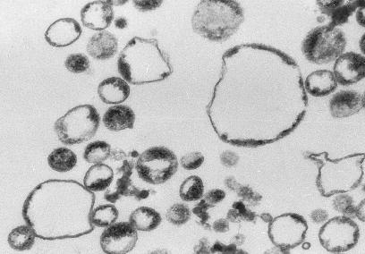
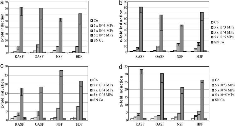
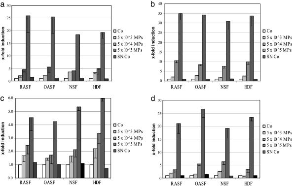
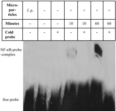
Rheumatoid arthritis is a chronic inflammatory disease characterized by destruction of cartilage and bone that is mediated by synovial fibroblasts. To determine the mechanisms by which these cells are activated to produce matrix metalloproteinases (MMPs), the effects of microparticles were investigated. Microparticles are small membrane-bound vesicles whose release from immune cells is increased during activation and apoptosis. Because microparticles occur abundantly in the synovial fluid in rheumatoid arthritis, they could represent novel stimulatory agents. Microparticles derived from T cells and monocytes strongly induced the synthesis of MMP-1, MMP-3, MMP-9, and MMP-13 in fibroblasts. The induction was time-dependent, with effects primarily observed after 36 h; under these conditions, MMP-2, MMP-14, and tissue inhibitor of MMP-1 (TIMP-1), TIMP-2, and TIMP-3 were not induced. Microparticles also increased the synthesis of inflammatory mediators including IL-6, IL-8, monocyte chemoattractant protein 1 (MCP-1), and MCP-2. In Ikappa-B-transfected synovial fibroblasts, MMPs were less inducible by microparticles compared with wild-type fibroblasts. Blocking of TNFalpha and IL-1beta with antibodies against TNFalpha and with IL-1 receptor antagonist did not abrogate stimulation by microparticles. These data provide evidence for a novel mechanism by which vesicles derived from activated or apoptotic immune cells can promote the destructive activity of synovial fibroblasts in rheumatoid arthritis.


Figures

Fig. 1.
Fig. 1.
Ultrastructural analysis of microparticles. Analysis of microparticles from Jurkat cells by transmission electron microscopy with an 80,000-fold magnification showed membrane-coated circular vesicles ranging in size from 200 to 700 nm.
Fig. 2.
Fig. 2.
Up-regulation of MMP-1 (a), MMP-3 (b), MMP-9 (c), and MMP-13 (d) mRNA in fibroblasts by microparticles from Jurkat cells. Cultured RASF, OASF, NSF, and HDFs were stimulated with increasing amounts of microparticles for 36 h. A strong induction of MMP-1, MMP-3, MMP-9, and MMP-13 was observed in all types of fibroblasts. Co, control; SN Co, supernatant control (see The Effects of Microparticles from Apoptotic and Activated Jurkat Cells on the Induction of MMP mRNA in Synovial Fibroblasts).
Fig. 3.
Fig. 3.
Induction of IL-6 (a), IL-8 (b), MCP-1 (c), and MCP-2 (d) by microparticles derived from Jurkat cells. These proinflammatory cytokines were up-regulated by microparticles. Comparable results were obtained with RASF, OASF, NSF, and NDF. Co, control; SN Co, supernatant control.
Fig. 4.
Fig. 4.
EMSA demonstrates the activation of NF-κB in RASF after stimulation with microparticles. Lane 1, free probe without nuclear extracts; lane 2, nuclear extract from unstimulated RASF, with NF-κB probe; lane 3, nuclear extract from unstimulated RASF, with NF-κB probe and cold NF-κB probe; lane 4, nuclear extract from RASF stimulated with microparticles for 10 min, with NF-κB probe; lane 5, nuclear extract from RASF stimulated with microparticles for 10 min, with NF-κB probe and cold NF-κB probe; lane 6, nuclear extract from RASF stimulated with microparticles for 60 min, with NF-κB probe; lane 7, nuclear extract from RASF stimulated with microparticles for 60 min, with NF-κB probe and cold NF-κB probe. Signals for NF-κB–DNA complexes in lane 4 and especially lane 6 confirm an activation of the transcription factor NF-κB. No activation of NF-κB signaling was observed in nonstimulated, serum-starved fibroblasts (lanes 2 and 3). The signal of the NF-κB–DNA complexes disappeared completely after adding an excess of unlabeled cold NF-κB probe (lanes 5 and 7).

References

    1. Firestein, G. S. (2003) Nature 423, 356–361. - PubMed
    1. Pap, T., Muller-Ladner, U., Gay, R. E. & Gay, S. (2000) Arthritis Res. 2, 361–367. - PMC - PubMed
    1. Vincenti, M. P. & Brinckerhoff, C. E. (2002) Arthritis Res. 4, 157–164. - PMC - PubMed
    1. Combes, V., Simon, A. C., Grau, G. E., Arnoux, D., Camoin, L., Sabatier, F., Mutin, M., Sanmarco, M., Sampol, J. & Dignat-George, F. (1999) J. Clin. Invest. 104, 93–102. - PMC - PubMed
    1. Zwaal, R. F. & Schroit, A. J. (1997) Blood 89, 1121–1132. - PubMed

MeSH terms