. 2005 Feb 15;102(7):2402-7.
doi: 10.1073/pnas.0409741102. Epub 2005 Feb 8.

Assembly and regulation of a glycolytic enzyme complex on the human erythrocyte membrane

Affiliations

Assembly and regulation of a glycolytic enzyme complex on the human erythrocyte membrane

M Estela Campanella et al. Proc Natl Acad Sci U S A. .

Abstract

To characterize the location of glycolytic enzymes (GEs) in intact human erythrocytes, freshly drawn blood was fixed and stained with Abs to GAPDH, aldolase, phosphofructokinase (PFK), pyruvate kinase (PK), lactate dehydrogenase (LDH), carbonic anhydrase II, Hb, and band 3 (AE1). Confocal microscopy revealed that in cells where band 3 displays its expected membrane staining and Hb is evenly distributed across the cytoplasm, GEs are largely limited to the membrane. Biochemical studies confirmed that the membrane binding sites for GAPDH, aldolase, and PFK reside on band 3, but related analyses demonstrate that sites for PK and LDH do not. Four lines of evidence demonstrate that the GEs are at least partially assembled into multimeric complexes near the NH2 terminus of band 3. First, a mAb to residues 1-12 of band 3 displaces all of the above GEs from the membrane, including LDH and PK, which do not bind band 3. Second, tyrosine phosphorylation of the NH2 terminus of band 3 (Y8 and Y21) reversibly releases all of the GEs from the membrane, including LDH and PK. Third, deoxygenation of RBCs dislodges all GEs from the membrane, consistent with the established ability of deoxyHb but not oxyHb to bind the NH2 terminus of band 3. Fourth, a large increase in the accessibility of enzyme epitopes is observed upon dissociation of GEs from the membrane. We conclude, therefore, that GEs are organized into complexes on the membrane whose assembly is regulated by oxygenation and phosphorylation.


Figures

Fig. 1.
Fig. 1.
Confocal microscopy of fresh human RBCs stained for aldolase, Hb, and band 3. Freshly drawn RBCs were processed as described in Materials and Methods, where the same sample was stained initially for Hb, then aldolase and finally band 3 (A) or initially for band 3 and then aldolase (B). (B) An overlay of aldolase and band 3 is provided to demonstrate the coincidence of their staining patterns.
Fig. 2.
Fig. 2.
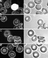
Confocal immunofluorescence (Left) and corresponding brightfield (Right) images of freshly drawn RBCs stained with Abs for GAPDH (A), PFK (B), LDH (C), and PK (D). Note that the only cell that displays strong cytoplasmic staining has spherocytic morphology. Additional micrographs and numerical analyses of cells with membrane localized enzymes are provided in Fig. 6 and Table 2.
Fig. 3.
Fig. 3.
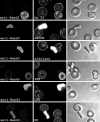
Impact of a mAb to the NH2 terminus of band 3 upon GE binding to the red-cell membrane. Washed RBCs were lysed and resealed in the presence of a mAb to residues 1–12 of band 3, as described in Materials and Methods. Entrapment of anti-band 3 (Left) was visualized by staining with donkey anti-mouse conjugated with Cy2, whereas enzyme localization in the same field of cells (Center) was visualized as in Fig. 2. (Right) Micrographs displaying a brightfield image of the same cells. Note that enzyme displacement from the membrane is seen only in cells that contain entrapped mAb. See Fig. 7 for additional micrographs.
Fig. 4.
Fig. 4.
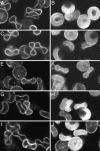
Effect of deoxygenation on the distribution of GEs in intact RBCs. Washed cells were deoxygenated by evacuation under argon and then fixed and stained as described in Materials and Methods.(A, C, E, G, and I) Samples equilibrated in air. (B, D, F, H, and J) Deoxygenated samples equilibrated in argon. Cells are stained for GAPDH (A and B), aldolase (C and D), PFK (E and F), LDH (G and H), and PK (I and J).
Fig. 5.
Fig. 5.
Analysis of pervanadate-induced tyrosine phosphorylation of human RBCs by immunoblotting and confocal microscopy. Freshly drawn RBCs were stimulated with pervanadate, as described in Materials and Methods. (A) Membranes were prepared, separated by SDS/PAGE, and analyzed by immunoblotting first with a monoclonal anti-phosphotyrosine IgG (Left) and, after stripping, with a monoclonal anti-band 3 (Right). Lanes: 1, untreated control; 2, RBCs treated with pervanadate (see Materials and Methods); 3, RBCs treated first with staurosporine to block tyrosine phosphorylation and then with pervanadate as in lane 2. (Left) A Coomassie blue-stained lane of ghosts is aligned next to the blot. (B) RBCs stimulated with pervanadate as in A were fixed and triply stained with anti-phosphotyrosine (Top), anti-GAPDH (Middle), and anti-LDH (Bottom) before imaging by confocal microscopy. Micrographs of untreated control cells (Left), pervanadate-treated cells (Center), and staurosporine plus pervanadate-treated cells (Right) are shown. Note that the magnitude of enzyme displacement is proportional to the magnitude of tyrosine phosphorylation (see also Fig. 8).

References

    1. Schrier, S. L. (1966) Am. J. Physiol. 210, 139-145. - PubMed
    1. Tanner, M. J & Gray, W. R. (1971) Biochem. J. 125, 1109-1117. - PMC - PubMed
    1. Tsai, I. H., Murthy, S. N. & Steck, T. L. (1982) J. Biol. Chem. 257, 1438-1442. - PubMed
    1. Murthy, S. N., Liu, T., Kaul, R. K., Kohler, H. & Steck, T. L. (1981) J. Biol. Chem. 256, 11203-11208. - PubMed
    1. Higashi, T., Richards, C. S. & Uyeda, K. (1979) J. Biol. Chem. 54, 9542-9550. - PubMed

Publication types

MeSH terms

Substances