. 2005 Feb 22;102(8):2766-71.
doi: 10.1073/pnas.0409719102. Epub 2005 Feb 8.

The association of epsin with ubiquitinated cargo along the endocytic pathway is negatively regulated by its interaction with clathrin

Affiliations

The association of epsin with ubiquitinated cargo along the endocytic pathway is negatively regulated by its interaction with clathrin

Hong Chen et al. Proc Natl Acad Sci U S A. .

Abstract

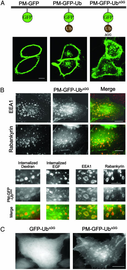
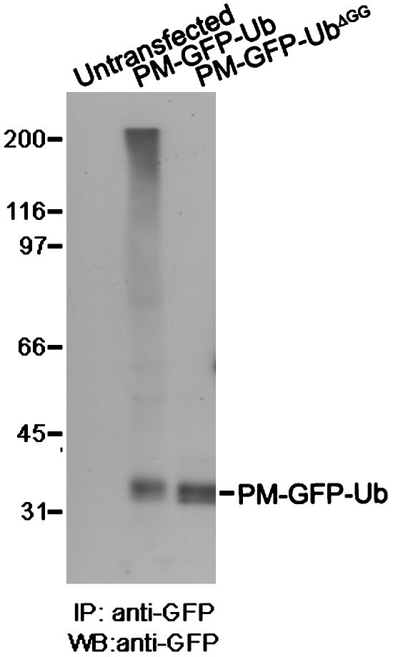
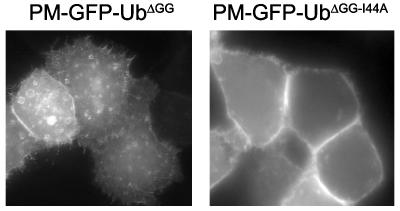
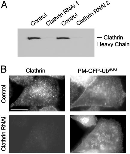
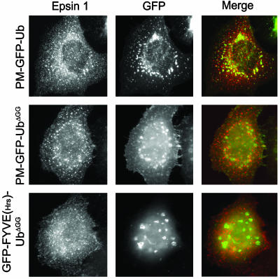
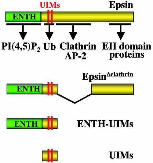
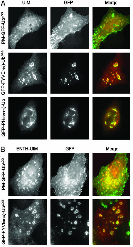
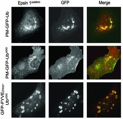
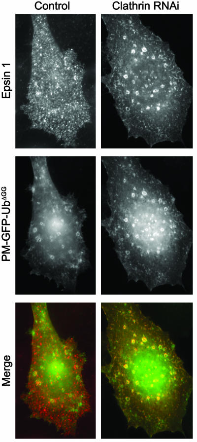
Monoubiquitination of plasma membrane proteins is a mechanism to control their endocytic trafficking by promoting their interaction with cytosolic adaptor proteins that contain ubiquitin (Ub)-binding domains. Epsin, which contains Ub interaction motifs (UIMs), as well as binding sites for the clathrin coat and clathrin accessory factors, is thought to function as one of such adaptors. The importance of clathrin in the internalization of ubiquitinated cargo, however, has been questioned. Here, we show that a GFP-Ub chimera directly targeted to the plasma membrane via a lipid-based interaction is efficiently taken up by endocytosis and delivered to the same endosomes that accumulate internalized EGF. Internalization of the chimera requires integrity of the UIM binding interface of Ub, but does not require clathrin. Surprisingly, WT epsin showed little colocalization with this chimera, whereas UIM-containing epsin constructs that lack the clathrin and AP2 binding region, strikingly colocalized with this chimera on endocytic vacuoles. In addition, extensive colocalization of WT epsin with the chimera on endocytic structures could be observed in cells where clathrin levels were drastically reduced by RNA interference. Our results reveal an important regulatory mechanism in epsin function. The mutually exclusive colocalization of epsin with membrane-bound Ub or clathrin may play a role in controlling the endocytic route taken by ubiquitinated cargo.


Figures

Fig. 1.
Fig. 1.
PM-GFP is selectively localized at the PM, whereas PM-GFP-Ub and PM-GFP-UbΔGG are internalized and also accumulate on intracellular endocytic compartments. (A) Confocal microscopy images of HeLa cells. (B) HeLa cells transfected with PM-GFP-UbΔGG were incubated with or without Alexa Fluor 594-conjugated dextran or rhodamine-conjugated EGF, then fixed and either processed by immunofluorescence for the endosomal proteins EEA1 and rabankyrin or examined directly for the internalized dye-conjugated endocytic probes. (C) Epifluorescence microscopy of HeLa cells transfected with the constructs indicated. Note the diffuse cytosolic localization of GFP-UbΔGG that lacks the short amino acid sequence directing acylation and PM localization. (Bars: 12 μm.)
Fig. 2.
Fig. 2.
PM-GFP-Ub, but not PM-GFP-UbΔGG, is incorporated into polyubiquitin chains. Anti-GFP immunoprecipitates from detergent extracts of untransfected cells and cells transfected with GFP constructs were processed by Western blotting for GFP immunoreactivity. IP, immunoprecipitation; WB, Western blot.
Fig. 3.
Fig. 3.
The internalization of PM-GFP-UbΔGG is abolished by the substitution of isoleucine 44 with alanine as examined by epifluorescence microscopy of HeLa cells. (Magnification: ×500.)
Fig. 4.
Fig. 4.
Endocytosis of PM-GFP-Ub does not depend on clathrin. (A) Anticlathrin Western blot of extracts from cells treated with control siRNA and two distinct sets of clathrin siRNA duplexes. (B) HeLa cells treated with control siRNA or a mixture of the two siRNA duplexes used for A were transfected with PM-GFP-UbΔGG, then fixed and analyzed for GFP fluorescence and clathrin immunoreactivity. (Bar: 12 μm.)
Fig. 5.
Fig. 5.
Full-length epsin 1 shows little colocalization with GFP-Ub constructs. CHO cells (Top) or HeLa cells (Middle and Bottom) cotransfected with epitope-tagged full-length epsin 1 and GFP-Ub constructs were fixed and examined for GFP fluorescence and epitope tag immunoreactivity by epifluorescence microscopy. Note the typical punctate pattern of epsin, reflecting clathrin-coated pits localization (5), regardless of the subcellular localization of GFP-Ub constructs. (Magnification: ×750.)
Fig. 6.
Fig. 6.
Domain cartoon of epsin and epsin constructs used in this study. Interacting regions for binding partners are indicated by arrows.
Fig. 7.
Fig. 7.
Fragments of epsin containing the UIM (A) or the ENTH-UIM (B) region colocalize extensively with internalized PM-GFP-UbΔGG. HeLa cells cotransfected with GFP-Ub constructs and epitope-tagged UIMs or ENTH-UIMs were fixed and examined for GFP fluorescence and epitope tag immunoreactivity. Note colocalization of UIMs and ENTH-UIMs with GFP-Ub constructs irrespective of their subcellular localization. (Magnification: ×800.)
Fig. 8.
Fig. 8.
An epitope-tagged epsin mutant (EpsinΔclathrin) lacking the clathrin and AP-2 binding region colocalized with PM-GFP-UbΔGG and GFP-FYVE(Hrs)-UbΔGG on endocytic compartments. Cotransfected CHO cells (Top) or HeLa cells (Middle and Bottom) were fixed and examined for GFP fluorescence and epitope tag immunoreactivity. (Magnification: ×750.)
Fig. 9.
Fig. 9.
A pool of full-length epsin colocalizes with PM-GFP-UbΔGG on intracellular endocytic vacuoles after RNAi-mediated clathrin knockdown. HeLa cells pretreated with control siRNA or clathrin heavy chain-specific siRNA duplexes were transfected with epitope-tagged full-length epsin 1 construct and PM-GFP-UbΔGG. Cells were then fixed and analyzed for GFP fluorescence and epitope tag immunoreactivity. (Magnification: ×1,000.)

Comment in

References

    1. Hershko, A. & Ciechanover, A. (1998) Annu. Rev. Biochem. 67, 425–479. - PubMed
    1. Hicke, L. & Dunn, R. (2003) Annu. Rev. Cell. Dev. Biol. 19, 141–172. - PubMed
    1. Pickart, C. M. (2001) Annu. Rev. Biochem. 70, 503–533. - PubMed
    1. Aguilar R. C. & Wendland, B. (2003) Curr. Opin. Cell Biol. 15, 184–190. - PubMed
    1. Chen, H., Fre, S., Slepnev, V. I., Capua, M. R., Takei, K., Butler, M. H., Di Fiore, P. P. & De Camilli, P. (1998) Nature 394, 793–797. - PubMed

Publication types