. 2005 Feb 15;102(7):2373-8.
doi: 10.1073/pnas.0409572102. Epub 2005 Feb 8.

Symmetry and frustration in protein energy landscapes: a near degeneracy resolves the Rop dimer-folding mystery

Affiliations

Symmetry and frustration in protein energy landscapes: a near degeneracy resolves the Rop dimer-folding mystery

Yaakov Levy et al. Proc Natl Acad Sci U S A. .

Abstract

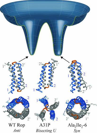
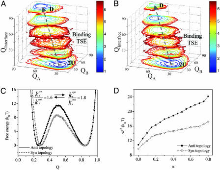
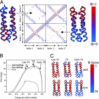
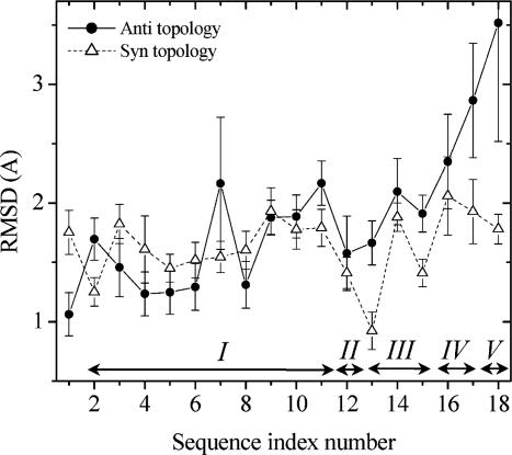
Protein folding has become one of the best understood biochemical reactions from a kinetic viewpoint. The funneled energy landscape, a consequence of the minimal frustration achieved by evolution in sequences, explains how most proteins fold efficiently and robustly to their functional structure and allows robust prediction of folding kinetics. The folding of Rop (repressor of primer) dimer is exceptional because some of its mutants with a redesigned hydrophobic core both fold and unfold much faster than the WT protein, which seems to conflict with a simple funneled energy landscape for which topology mainly determines the kinetics. We propose that the mystery of Rop folding can be unraveled by assuming a double-funneled energy landscape on which there are two basins that correspond to distinct but related topological structures. Because of the near symmetry of the molecule, mutations can cause a conformational switch to a nearly degenerate yet distinct topology or lead to a mixture of both topologies. The topology predicted to have the lower free-energy barrier height for folding was further found by all-atom modeling to give a better structural fit for those mutants with the extreme folding and unfolding rates. Thus, the non-Hammond effects can be understood within energy-landscape theory if there are in fact two different but nearly degenerate structures for Rop. Mutations in symmetric and regular structures may give rise to frustration and thus result in degeneracy.


Figures

Fig. 1.
Fig. 1.
A schematic representation of a double-funneled energy landscape of Rop dimer and its three structural topologies that correspond to the WT sequence and the mutants Ala2Ile2-6 and A31P. In these structures, one monomer is colored gray, and the other monomer is colored blue. The loop between the two helices in each monomer is colored orange. Residues Lys-3, Asn-10, Gln-18, and Lys-25 in helices 1 and 1′, which constitute the binding site to the RNA (39), are shown by stick representation. The structure of WT Rop dimer (11) and the Ala2Ile2-6 mutant (34) is a four-helix coiled coil and has been determined by x-ray crystallography [the structure of the WT Rop was also assigned by NMR (12)]. The WT Rop has an anti topology and the Ala2Ile2-6 has a syn topology, which is obtained by a 180° flip of one monomer around an axis normal to the dimer interface. The mutation Ala-31 into Pro is located in the loop and introduces a conformational change into a bisecting U topology (16). The anti and syn topologies are nearly degenerate and correspond to the two basins. The bisecting U is expected to be less stable than the anti and syn topologies and was not placed on the double-funneled energy landscape because its folding was not studied here.
Fig. 2.
Fig. 2.
The barrier for the folding of the anti and syn forms of Rop dimer. The folding free-energy landscapes for the anti (A) and syn (B) topologies of Rop dimer are shown. The reaction coordinates are the folding of the two monomers and the formation of the interface (i.e., association). U, an unfolded monomer; D, a folded dimer. The dashed arrow illustrates the coupling between folding and association. (C). Two-dimensional free-energy profiles for the folding and association of the two forms of the Rop dimer based on the additive native topology-based simulations. The rates for folding and unfolding for each topological structure were obtained from >1,000 events (using the additive model) that were fitted to a single exponential decay. (D) The folding barrier height, ΔF#, as a function of α (the three-body contribution to the contact energy).
Fig. 3.
Fig. 3.
The structure of the binding TSE of the Rop topologies. (A) Φ value analysis for the anti and syn topologies of Rop dimer. The contact and residue Φ values for the anti (upper triangle) and syn (lower triangle) forms of the Rop proteins. The contact Φ values, φij, for each native contact pair between i and j is calculated from the probability of contact formation, formula image. For Rop dimer, the superscripts F, U, and TS correspond to the folded dimer, unfolded monomers, and folding TSEs, respectively. The φi value of residue i is calculated from the contact values, φij, by averaging all of the φij values that are involved with residue i. The residue Φ values are presented by showing the corresponding residues in each topology in blue and red, corresponding to Φ equals to 0 and 1, respectively. (B) Free-energy profiles of Rop topologies. The profile of a typical single-domain protein U1A is also shown as a comparison. The x axis is normalized by the total number of contacts. The y axis is in the unit of monomer contact energy and is in approximate kilocalories/mole. (C) The B factor calculated for six important points along the two folding pathways (labeled by arrows in B).
Fig. 4.
Fig. 4.
The average rmsd of each designed Rop mutant as anti and syn topologies in respect to the x-ray structures of the WT and Ala2Ile2-6 mutants. Each designed structure was simulated with all-atom representation of the protein with explicit solvent model for 5 ns. To account for different packing of the two monomers, the rmsd was calculated after superimposing a single monomer. The arrows indicate the mutant classes as in Table 1.

References

    1. Onuchic, J. N., Luthey-Schulten, Z. & Wolynes, P. G. (1997) Annu. Rev. Phys. Chem. 48, 539-594. - PubMed
    1. Onuchic, J. N. & Wolynes, P. G. (2004) Curr. Opin. Struct. Biol. 14, 70-75. - PubMed
    1. Gunasekaran, K., Eyles, S. J., Hagler, A. T. & Gierasch, L. M. (2001) Curr. Opin. Struct. Biol. 2001, 83-93. - PubMed
    1. Murzin, A., Brenner, S., Hubbard, T. & Chothia, C. (1995) J. Mol. Biol. 247, 536-540. - PubMed
    1. Orengo, C., Jones, D. & Thornton, J. (1994) Nature 372, 631-634. - PubMed

Publication types

Substances

LinkOut - more resources