. 2005 Jun 1;105(11):4345-52.
doi: 10.1182/blood-2004-07-2718. Epub 2005 Feb 8.

The beta3 subunit of the integrin alphaIIbbeta3 regulates alphaIIb-mediated outside-in signaling

Affiliations

The beta3 subunit of the integrin alphaIIbbeta3 regulates alphaIIb-mediated outside-in signaling

Junling Liu et al. Blood. .

Abstract

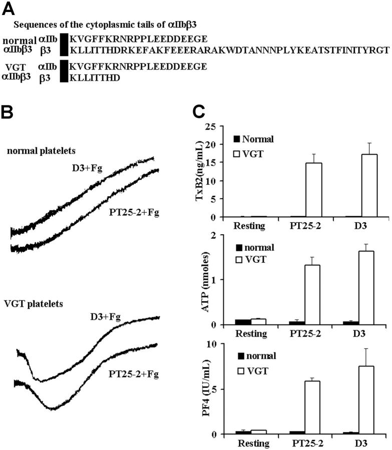
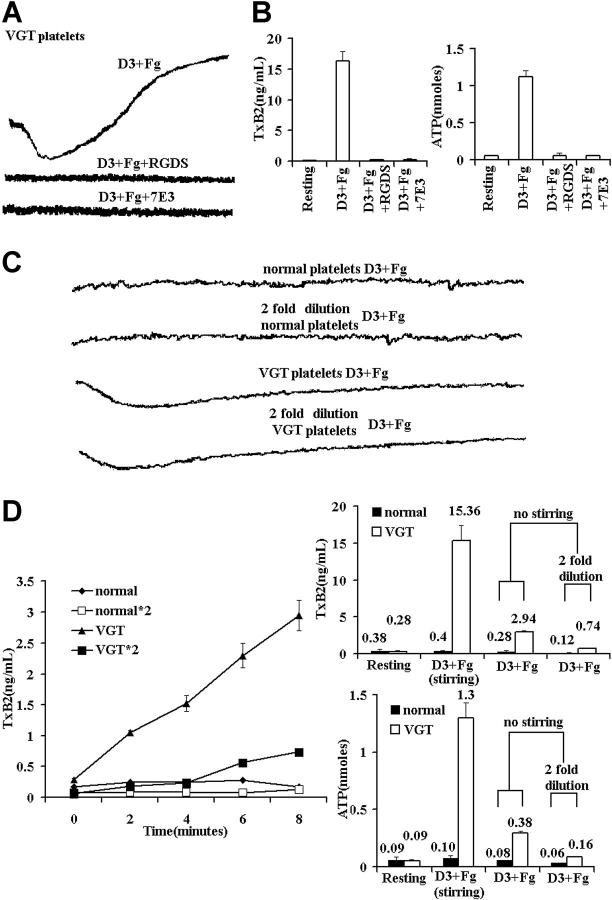
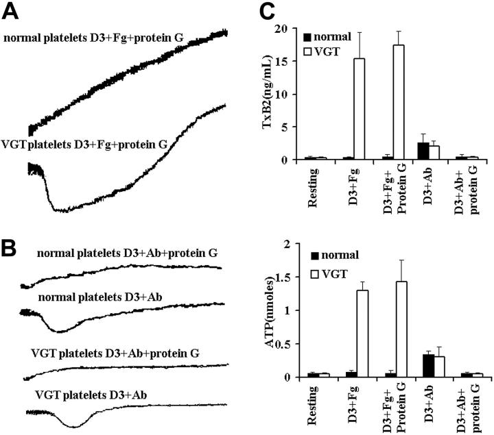
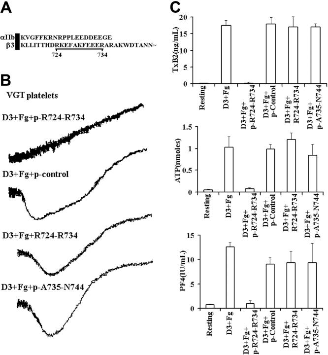
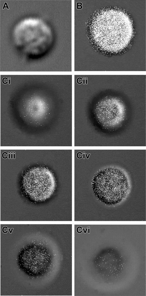
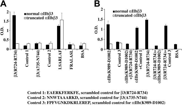
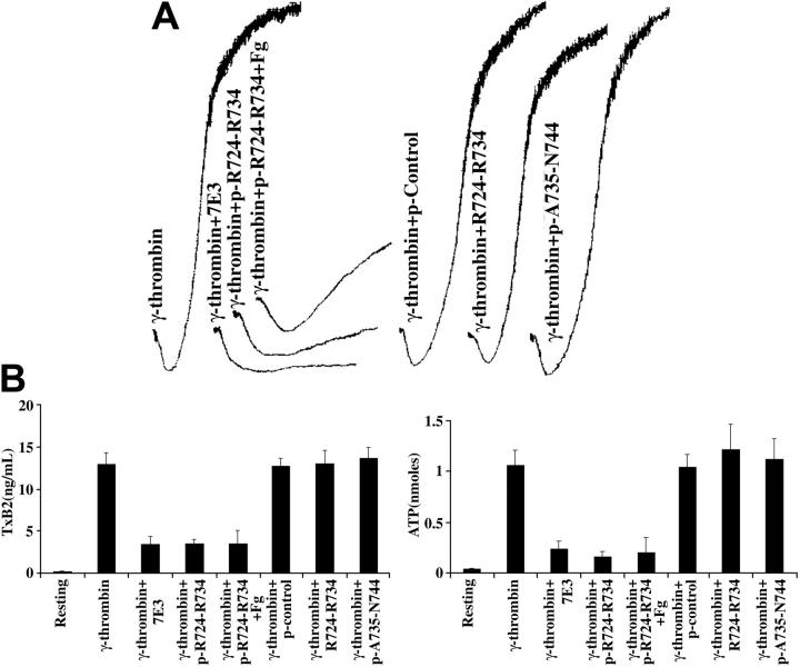
Bidirectional signaling is an essential feature of alphaIIbbeta3 function. The alphaIIb cytoplasmic domain negatively regulates beta3-mediated inside-out signaling, but little is known about the regulation of alphaIIb-mediated outside-in signaling. We show that alphaIIb-mediated outside-in signaling is enhanced in platelets of a patient lacking the terminal 39 residues of the beta3 cytoplasmic tail. This enhanced signaling was detected as thromboxane A(2) (TxA(2)) production and granule secretion, and required ligand cross-linking of alphaIIbbeta3 and platelet aggregation. This outside-in signaling was specifically inhibited by a palmitoylated version of a beta3 peptide corresponding to cytoplasmic domain residues R724-R734. Unlike the palmitoylated peptide, the nonpalmitoylated beta3 peptide could not cross the platelet membrane and did not inhibit this outside-in signaling. The physiologic relevance of this beta3-mediated negative regulation of alphaIIb outside-in signaling was demonstrated in normal platelets treated with the palmitoylated peptide and a physiologic agonist. Binding of alphaIIbbeta3 complexes to immobilized peptides demonstrated that a peptide corresponding to beta3 residues R724-R734 appears to bind to an alphaIIb cytoplasmic domain peptide containing residues K989-D1002, but not to control peptides. These results demonstrate that alphaIIb-mediated outside-in signaling resulting in TxA(2) production and granule secretion is negatively regulated by a sequence of residues in the membrane distal beta3 cytoplasmic domain sequence RKEFAKFEEER.


Figures

Figure 1.
Figure 1.
LIBS-specific mAb-induced signaling by platelets lacking the β3 cytoplasmic domain. (A) Sequences of the αIIb and β3 cytoplasmic domains present in normal and VGTΔ724 platelets. (B) Aggregation traces of normal and VGTΔ724 platelets treated separately with the LIBS-specific mAbs D3 (30 μg/mL) and PT25-2 (30 μg/mL) in the presence of fibrinogen (Fg; 250 μg/mL). It is noteworthy that D3 and PT25-2 each induced shape change of VGTΔ724, but not the normal platelets. (C) Graph presentation of TxA2 production as well as ATP and PF4 secretion. The error bars represent SD, n = 3. In some cases, the values of the SD were so small that the bars cannot be seen.
Figure 2.
Figure 2.
αIIb-mediated outside-in signaling requires fibrinogen binding and aggregation. (A) Aggregation traces of VGTΔ724 platelets treated with D3 (30 μg/mL) in the presence of Fg (250 μg/mL), with or without the peptide RGDS (1 mM) or the mAb 7E3 (10 μg/mL) (RGDS and 7E3 prevent Fg binding to αIIbβ3). (B) D3 plus Fg-induced TxA2 production and ATP secretion by VGTΔ724 platelets treated with substances that inhibit Fg binding to αIIbβ3. (C) Aggregation traces of (450 μL) normal and VGTΔ724 platelets in response to D3 (30 μg/mL) plus Fg (250 μg/mL) without stirring. (D) TxA2 production by normal and VGTΔ724 platelets was measured at zero time and every 2 minutes for 8 minutes; the final concentration of secreted ATP also was measured. Data shown here were from 3 experiments. The error bars represent SD, n = 3. In some cases, the values of the SD were so small that the bars cannot be seen.
Figure 3.
Figure 3.
The signaling induced by the LIBS-specific antibodies is not Fc receptor (FcγRIIA)–dependent. (A) Aggregation traces of normal and VGTΔ724 platelets treated with D3 (30 μg/mL) plus Fg (250 μg/mL) in the presence of protein G (15 μg/mL). (B) Aggregation traces of normal and VGTΔ724 platelets treated with D3 (30 μg/mL) plus donkey anti–mouse polyclonal antibody (Ab; 30 μg/mL) with or without protein G. (C) TxA2 production (top) and ATP secretion (bottom) by normal and VGTΔ724 platelets in response to different treatments which inhibit or enhance Fc receptor–mediated signaling. Data shown here were from 3 experiments. Error bars represent SD, n = 3. In some cases, the values of the SD were so small that the bars cannot be seen.
Figure 4.
Figure 4.
The β3 palmitoylated peptide pRKEFAKFEEER (pR724-R734) negatively regulates αIIb-mediated outside-in signaling. (A) Sequences of the αIIb and β3 cytoplasmic domains present in normal platelets. (B) Aggregation traces of VGTΔ724 platelets treated with D3 (30 μg/mL) in the presence of Fg (250 μg/mL) with or without the β3 palmitoylated peptide p-R724-R734 (pRKEFAKFEEER, 10 μM), which corresponds to the region of β3 underlined in panel A; this sequence is part of the cytoplasmic domain missing in VGTΔ724 platelets. The peptides pEAERKFERKFE (p control; 10 μM), a scrambled, palmitoylated version of pRKEFAKFEEER, a nonpalmitoylated form of the peptide R724-R734 (RKEFAKFEEER, 10 μM), and the palmitoylated β3 peptide p-A735-N744 (pARAKWDTANN) were used as controls. In contrast to the control peptides, pRKEFAKFEEER eliminated secretion-induced shape change by D3 plus Fg-treated VGTΔ724 platelets. (C) TxA2 production (top) as well as ATP (middle) and PF4 secretion (bottom) by VGTΔ724 platelets treated with D3 (30 μg/mL) in the presence of Fg (250 μg/mL) with or without p-R724-R734 (10 μM), or 10 μM of the control peptides. There are no significant differences (P < .05) between the values of D3 plus Fg, D3 plus Fg plus pEAERKFERKFE or pARAKWDTANN-treated platelets for TxA2 production and secretion of ATP and PF4. The error bars represent SD, n = 4. In some cases, the values of the SD were so small that the bars cannot be seen.
Figure 5.
Figure 5.
The β3 palmitoylated peptide pRKEFAKFEEER but not the nonpalmitoylated version RKEFAKFEEER is platelet permeable. (A) A confocal projection image composed of optical slice images of a single representative normal platelet treated with the FITC-derivatized, nonpalmitoylated peptide RKEFAKFEEE. Images of optical slices of the platelet treated with this peptide revealed no label (not shown). (B) A confocal projection image composed of the optical slice images of single representative normal human platelet treated with the FITC-derivatized, palmitoylated peptide pRKEFAKFEEER. (C) Six representative noncontiguous consecutive optical slice images of this platelet are labeled i-vi. (i) An image of the ventral surface; (ii-v) images of the interior of the platelets; (vi) an image of the dorsal surface of the platelet. The white points in the images represent the FITC fluorescence. These images demonstrate that palmitoylated RKEFAKFEEER, but not nonpalmitoylated RKEFAKFEEER, is platelet permeable and therefore can enter the platelets and reside in their interior.
Figure 6.
Figure 6.
The β3 cytoplasmic domain peptide RKEFAKFEEER binds to the cytoplasmic domain of αIIb. The details of the methodology for the binding experiments are described in “Patient, materials, methods.” (A) Normal αIIbβ3 bound to the peptides RKEFAKFEEER and LSARLAF, but not to the β3 peptide ARAKWDTANN, or the scrambled control peptides EAERKFERKFE, NNWTAAARKD, and FRALASL. In contrast, truncated αIIbβ3 did not bind to RKEFAKFEEER; it bound only to LSARLAF. (B) Normal αIIbβ3 bound to the αIIb cytoplasmic domain peptide KVGFFKRNRPPLEED but not to the scrambled version FPFVGNKDKRLEREP. Truncated αIIbβ3 did not bind to KVGFFKRNRPPLEED or FPFVGNKDKRLEREP. The β3 peptide RKEFAKFEEER but not its scrambled control version (Control 1) inhibited the binding of αIIbβ3 to immobilized KVGFFKRNRPPLEED. Conversely, KVGFFKRNRPPLEED, but not the scrambled control version (Control 3) inhibited the binding of αIIbβ3 to RKEFAKFEEER. The simplest interpretation of these data is that RKEFAKFEEER or a sequence therein can bind to the cytoplasmic domain of αIIb. The data represent the results of 3 experiments. The error bars represent SD, n = 3. In some cases, the values of the SD were so small that the bars cannot be seen.
Figure 7.
Figure 7.
Palmitoylated peptide pR724-R734 (pRKEFAKFEEER) inhibited low-level γ-thrombin–induced αIIbβ3-mediated outside-in signaling and its associated TxA2 production and ATP secretion. (A) Aggregation traces of normal platelets treated with low-level γ-thrombin (5 nM) in the presence of 7E3 (10 μg/mL), the palmitoylated peptide p-R724-R734 (pRKEFAKFEEER, 10 μM) with or without Fg (250 μg/mL), a scrambled, palmitoylated control peptide (pEAERKFERKFE, 10 μM), a nonpalmitoylated version of peptide R724-R734, (RKEFAKFEEER, 10 μM), or the palmitoylated peptide pA735-N744 (pARAKWDTANN, 10 μM). In contrast to pARAKWDTANN and the control peptides, 7E3 and pRKEFAKFEEER inhibited aggregation induced by low-level γ-thrombin. Exogenous Fg restored aggregation to platelets treated with pRKEFAKFEEER. (B) TxA2 production (left) and ATP secretion (right) by normal platelets treated with γ-thrombin alone or in the presence of 7E3 (10 μg/mL), pRKEFAKFEEER (10 μM) with or without Fg, or the control peptides (10 μM). There are no significant differences (P < .05) between the values of 7E3 and pRKEFAKFEEER with or without Fg-treated platelets for TxA2 production and ATP secretion. The error bars represent SD, n = 3.

Similar articles

Cited by

References

    1. Hynes RO. Integrins: bidirectional, allosteric signaling machines. Cell. 2002;110: 673-687. - PubMed
    1. Lefkovits J, Plow EF, Topol EJ. Platelet glycoprotein IIb/IIIa receptors in cardiovascular medicine. N Engl J Med. 1995;332: 1553-1559. - PubMed
    1. Shattil SJ, Kashiwagi H, Pampori N. Integrin signaling: the platelet paradigm. Blood. 1998;91: 2645-2657. - PubMed
    1. Wang R, Shattil SJ, Ambruso DR, Newman PJ. Truncation of the cytoplasmic domain of beta3 in a variant form of Glanzmann thrombasthenia abrogates signaling through the integrin alpha(IIb)-beta3 complex. J Clin Invest. 1997;100: 2393-2403. - PMC - PubMed
    1. Tadokoro S, Shattil SJ, Eto K, et al. Talin binding to integrin beta tails: a final common step in integrin activation. Science. 2003;302: 103-106. - PubMed

Publication types