. 2005 Jun 1;105(11):4390-8.
doi: 10.1182/blood-2004-11-4284. Epub 2005 Feb 8.

Identification and characterization of circulating human transitional B cells

Affiliations

Identification and characterization of circulating human transitional B cells

Gary P Sims et al. Blood. .

Abstract

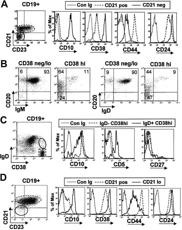
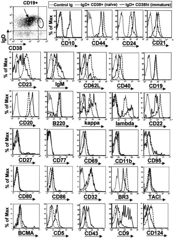
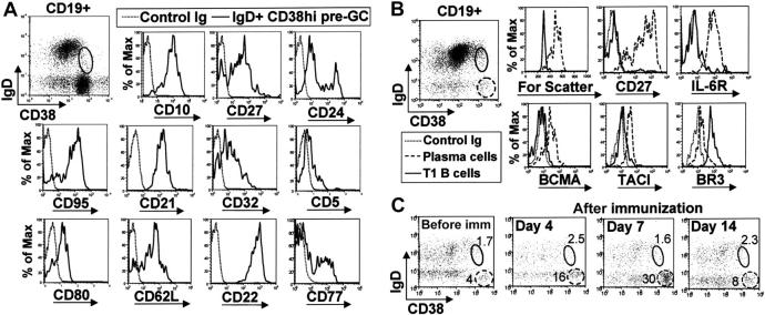
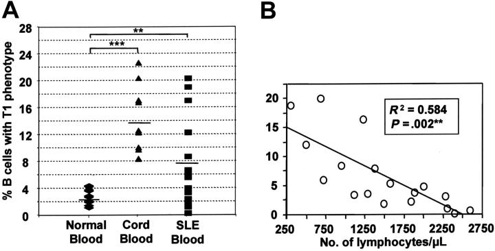
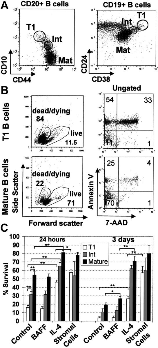
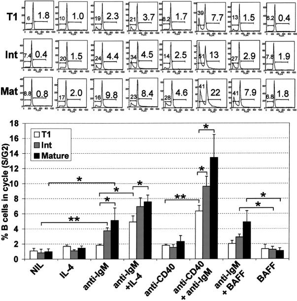
Murine B-cell development begins in bone marrow and results in the generation of immature transitional B cells that transit to the spleen to complete their maturation. It remains unclear whether the same developmental pathway takes place in humans. Using markers characteristic of human bone marrow immature B cells, we have identified a population of circulating human B cells with a phenotype most similar to mouse transitional type I (T1) B cells, although these human counterparts express CD5. These cells die rapidly in culture, and B-cell activation factor member of the tumor necrosis factor (TNF) family (BAFF) does not effect their survival regardless of B-cell receptor (BCR) stimulation. In contrast, bone marrow stromal cells or interleukin-4 (IL-4) significantly enhanced their survival. In the presence of T-cell signals provided by IL-4 or CD40 ligation, BCR stimulation can induce progression into cell cycle. Interestingly, circulating B cells that phenotypically and functionally resemble murine T2 B cells are found in cord blood and adult peripheral blood, suggesting that B-cell maturation may not be restricted to the spleen. Notably, increased proportions of T1 B cells were found in blood of patients with systemic lupus erythematosus (SLE), although bone marrow production and selection appeared to be normal.


Figures

Figure 1.
Figure 1.
Immature B-cell markers expressed by adult bone marrow and term cord-blood B cells. (A) Bone marrow CD19+ B cells were gated into CD21- immature and CD21+ mature fractions and the differential expression of CD10, CD38, CD44, and CD24 was assessed relative to isotype matched control mAb (Con Ig). (B) Bone marrow CD19+ B cells were gated into CD38neg/lo mature B cells and a CD38hi immature subset. A fraction of the latter express CD20 more densely and both IgM and IgD. (C) Differential expression of CD5 and CD10 by bone marrow IgD- CD38hi pro/pre B cells and IgD+ CD38hi immature B cells. (D) Cord blood CD19+ B cells were gated into CD21neg/lo and CD21+ fractions, and the differential expression of immature bone marrow markers was assessed. Results are representative of 2 bone marrow aspirates and 10 cord blood samples.
Figure 2.
Figure 2.
Circulating IgD+ CD38hi B cells express immature B-cell markers with an overall phenotype similar to transitional type I (T1) B cells. B cells were enriched from blood of healthy adult donors and stained with anti-CD19, anti-IgD, anti-CD38, and a fourth anti–human mAb. The differential expression of a variety of B-cell markers was compared between the IgD+ CD38hi immature population (solid line) and IgD+ CD38+ mature naive B cells (dashed line, indicated with arrow). Results are representative of data from at least 4 healthy adult donors.
Figure 3.
Figure 3.
Peripheral-blood IgD+ CD38hi T1 B cells are not circulating pre–germinal-center or plasma-cell precursors. (A) Tonsil mononuclear cells were stained with anti-CD19, anti-IgD, anti-CD38, and a fourth mAb to examine the phenotype of IgD+ CD38hi pre–germinal center B cells. (B) Enriched B cells from peripheral blood of a healthy donor were similarly stained to examine the expression of plasma cell markers on CD19+ IgD+ CD38hi T1 B cells (solid gate) and CD19lo IgD- CD38bright circulating plasma cells (dashed gate). (C) The relative frequencies of the CD19+ IgD+ CD38hi T1 B-cell population (solid gate) and CD19lo IgD- CD38bright plasma cells (dashed gate) from a healthy donor were examined before immunization and 4, 7, and 14 days after immunization with influenza vaccine.
Figure 4.
Figure 4.
T1 B cells are found in increased proportions in term cord blood and systemic lupus erythematosus patients. (A) The relative frequency of T1 B cells was determined for term cord blood (n=10), and the peripheral blood of healthy adult donors (n=29) and patients with systemic lupus erythematosus (SLE; n=18). Horizontal bars indicate means. Significant differences using the nonparametric Mann-Whitney U test were detected at P <.01 (**) and P <.001(***). (B) A linear regression plot shows that the frequency of B cells with a T1 B-cell phenotype is inversely proportional to the absolute number of peripheral lymphocytes.
Figure 5.
Figure 5.
“Intermediate” B cells are present in cord and peripheral blood. B cells from (A) peripheral and (B) cord blood were stained with various combinations of immature and T1 B-cell markers. The T1 population (solid gate), naive B cells (dashed gate), and “intermediate B cells” (arrow) are indicated.
Figure 6.
Figure 6.
T1 B cells are short-lived in culture although survival can be improved by coculture with mouse bone marrow stromal cells or IL-4 but not BAFF. (A) Negatively selected peripheral blood B cells from healthy adult donors were stained with anti-CD20, anti-CD10, and anti-CD44 or anti-CD19, anti-CD24, and anti-CD38. T1 B cell (CD20hi CD10hi CD44lo or CD19+ CD24hi CD38hi), mature naive (Mat) (CD20+ CD10- CD44hi or CD19+, CD24lo, CD38lo) and intermediate (Int) (CD20+ CD10lo CD44hi or CD19+ CD24int, CD38int) B-cell populations were sorted as shown. Postsorting analysis indicated that each population was more than 95% pure. (B) T1 and mature B-cell populations were cultured for 24 hours and stained with annexin V and 7-AAD. (C) T1 B cells, intermediate and mature B cell populations were cultured in medium alone or with the addition of either BAFF (200 ng/mL), IL-4 (100 ng/mL), or cultured on mouse S13 bone marrow stromal cells. After 24 hours or 3 days in culture, the cells were examined for viability by flow cytometry using forward/side scatter characteristics or 7-AAD exclusion. The data show the means ± SEM of at least 4 independent experiments. Unpaired t tests were used to detect significant differences (*P <.05; **P <.01).
Figure 7.
Figure 7.
T1 B cells have reduced capacity to enter cell cycle following B-cell receptor engagement. T1, mature, and intermediate B-cell populations (Figure 4A) were cultured alone (NIL) or stimulated with various combinations of IL-4 (100 ng/mL), anti-IgM (10 μg/mL), anti-CD40 (1 μg/mL), or BAFF (200 ng/mL) for 48 hours. Afterward, hypotonic propidium iodide staining was carried out to determine apoptotic and cycling cells. Forward/side scatter and FL2 width gating were used to gate out nuclear fragments and doublets. The FL2 area was examined to identify the frequency of sub G0/G1 apoptotic cells (left number) and S/G2 cycling cells (right number). A representative experiment is shown in the top panel, and the means ± SEM of 4 independent experiments is shown in the bottom panel. Unpaired t tests were used to detect significant differences (*P <.05; **P <.01).

Similar articles

Cited by

References

    1. Hartley SB, Crosbie J, Brink R, Kantor AB, Basten A, Goodnow CC. Elimination from peripheral lymphoid-tissues of self-reactive lymphocytes-B recognizing membrane-bound antigens. Nature. 1991;353: 765-769. - PubMed
    1. Nemazee DA, Burki K. Clonal deletion of lymphocyte-B in a transgenic mouse bearing anti-Mhc class-I antibody genes. Nature. 1989;337: 562-566. - PubMed
    1. Norvell A, Mandik L, Monroe JG. Engagement of the antigen-receptor on immature murine B-lymphocytes results in death by apoptosis. J Immunol. 1995;154: 4404-4413. - PubMed
    1. Forster I, Rajewsky K. The bulk of the peripheral B-cell pool in mice is stable and not rapidly renewed from the bone-marrow: Proc Natl Sci U S A. 1990;87: 4781-4784. - PMC - PubMed
    1. Loder F, Mutschler B, Ray RJ, et al. B cell development in the spleen takes place in discrete steps and is determined by the quality of B cell receptor-derived signals. J Exp Med. 1999;190: 75-89. - PMC - PubMed