. 2005 Mar;11(3):294-307.
doi: 10.1261/rna.5460105.

Perinuclear localization of slow troponin C m RNA in muscle cells is controlled by a cis-element located at its 3' untranslated region

Affiliations

Perinuclear localization of slow troponin C m RNA in muscle cells is controlled by a cis-element located at its 3' untranslated region

Kishore K Reddy et al. RNA. 2005 Mar.

Abstract

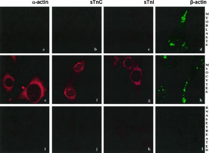
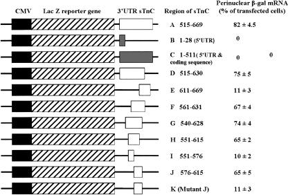
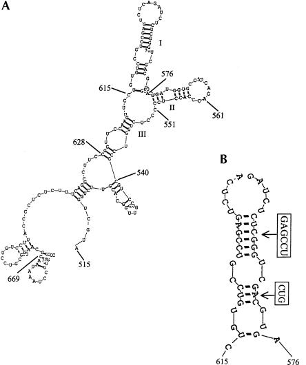
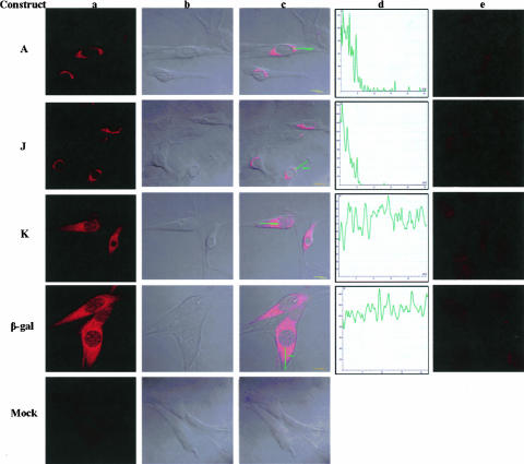
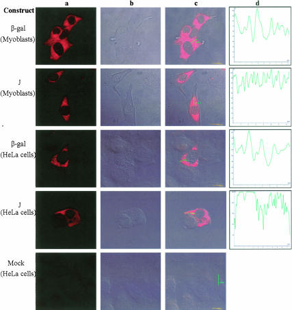
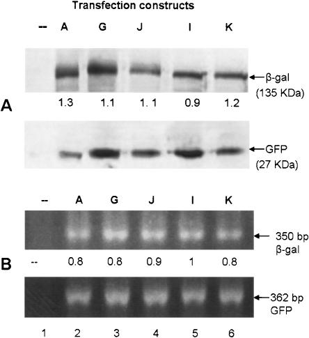
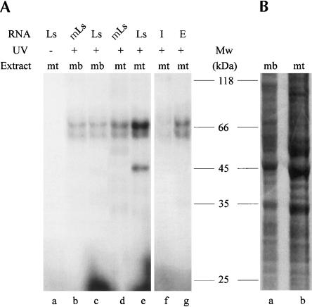
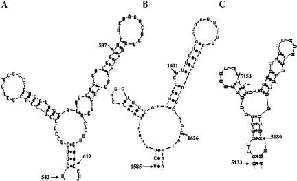
The process of mRNA localization within a specific cytoplasmic region is an integral aspect of the regulation of gene expression. Furthermore, colocalization of mRNAs and their respective translation products may facilitate the proper assembly of multi-subunit complexes like the thick and thin filaments of muscle. This postulate was tested by investigating the cytoplasmic localization of three mRNAs-the alpha-actin, slow troponin C (sTnC), and slow troponin I (sTnI), which encode different poly-peptide partners of the thin filament. Using in situ hybridization we showed that all three thin filament mRNAs are localized in the perinuclear cytoplasm of cultured C2C12 muscle cells. Their localization differs from that of the nonmuscle beta-actin mRNA, which is localized in the peripheral region of both proliferating nondifferentiated myoblasts and the differentiated myocytes. Analysis of the localization signal of the sTnC mRNA showed that a 40-nucleotide-long region of the sTnC mRNA 3' UTR is sufficient to confer the perinuclear localization on a heterologous reporter beta-Gal mRNA. This localization signal showed tissue specificity and worked only in the differentiated myocytes, but not in the proliferating myoblasts or in HeLa cells. The predicted secondary structure of the localization signal suggests the presence of multiple stem and loop structures in this region of the 3' UTR. Mutations within the stem region of the localization signal, which abolish the base pairing in this region, significantly reduced its perinuclear mRNA localization activity. Using UV-induced photo-cross-linking of RNA and proteins we found that a myotube-specific 42-kDa polypeptide binds to the localization signal.


Figures

FIGURE 1.
FIGURE 1.
Intracellular distribution of mRNAs. Mouse C2C12 cells were cultured on glass coverslips either as proliferating myoblasts or allowed to differentiate in the low-mitogen-containing medium. In situ hybridization with molecular beacon probes was performed, and specimens were examined by CSLM as described in Materials and Methods. Approximately 30–40 cells in 10 randomly chosen areas of the slide were viewed. The probes for detecting α-and β-actin mRNAs were labeled with CY3 and FAM, respectively. The probes for detecting sTnC and sTnI mRNAs were both labeled with TET. (a–d) In situ hybridization of myoblasts with α-actin (a), sTnC (b), sTnI (c), and β-actin (d) probes. Note the absence of strong signals for the muscle-specific mRNAs (a–c) in myoblasts and the presence of strong signals for nonmuscle β-actin mRNA. (e,h) In situ hybridization of differentiated myocytes that were maintained in the differentiation medium for 3 d. The strong hybridization signals for α-actin (e), sTnC (f), sTnI (g), and β-actin (h) can be seen. All three muscle-specific mRNAs show a concentration of signals around the nucleus, whereas the nonmuscle β-actin mRNA was concentrated near the leading edges in both myoblast (d) and myocytes (h). Fluorescent signal above the background was not detected when RNAse-treated specimens were hybridized to α-actin, β-actin, TnC, and TnI probes (i–l, respectively).
FIGURE 2.
FIGURE 2.
Distribution of β-Gal mRNA in transiently transfected C2C12 myocytes with β-gal-sTnC chimeras. These are schematic representations (not drawn to scale) of the different constructs used for β-Gal distribution studies. The CMV promoter (solid black box), and the Lac Z reporter gene (hatched box), the different segments of sTnC 3′ UTR sequences (open box), the 5′ untranslated region, and the coding region (gray box) are shown. Various regions of the sTnC mRNA were fused to the Lac Z reporter gene. Cells were transfected with each construct, and the cytoplasmic distribution of the β-Gal mRNA was analyzed by in situ hybridization. Approximately 50 transfected cells were examined, and the percentage of cells showing peri-nuclear localization was scored. The averages were calculated from six independent experiments.
FIGURE 3.
FIGURE 3.
The folding of the 3′ UTR of sTnC mRNA. The predicted secondary structure of (A) the entire 3′ UTR of sTnC mRNA excluding the last 18 nt containing the poly(A) addition signal, and (B) the 40-nt-long localization signal are shown. The structures were predicted using the RNA structure version 2.5 computer algorithm. The boxed nucleotides (B) indicate the base substitutions used in construct K (Fig. 2 ▶). The different stem and loop arrangements within nucleotides 540–628 have been designated as regions I, II, and III. The numbering refers to the nucleotide numbers of the sTnC mRNA beginning at its 5′ end (accession number AA656586).
FIGURE 4.
FIGURE 4.
Regulation of cytoplasmic distribution of β-Gal mRNA by the 3′ UTR of sTnC mRNA in differentiated muscle cells. Differentiating C2C12 cells were transfected with different β-Gal constructs as described in Materials and Methods. Twenty-four hours after transfection, the cells were fixed, and the cytoplasmic distribution of the β-Gal mRNA was examined by in situ hybridization. Cells transfected with constructs A and J (Fig. 2 ▶) show the perinuclear distribution of β-Gal mRNA. In contrast, cells transfected with the mutant zipcode containing construct K (Fig. 2 ▶) show a diffuse distribution of β-Gal mRNA. The parent pCMV-SPORT-β-Gal (β-Gal) transfected cells also show diffuse distribution of the β-Gal mRNA. Fluorescent signal is not visible in mock-transfected and RNAse-treated cells. The panels are as follows: (a) fluorescent signal of the Cy3-labeled hybridized probe; (b) phase contrast pictures (Nomarski) of the cells shown in panel a; (c) panels a and b are merged; (d) line scans of fluorescent cells in panel a using the Leica software; and (e) hybridization of RNAse-treated cells with the β-Gal molecular beacon probe.
FIGURE 5.
FIGURE 5.
Cell specificity of sTnC mRNA localization signal. Proliferating C2C12 myoblasts and HeLa cells were transfected with pCMV-SPORT-β-Gal or construct J containing the sTnC mRNAs 40-nt-long localization signal. Twenty-four hours after transfection, the cytoplasmic distribution of β-Gal mRNA was examined as described in Materials and Methods. (a) The diffuse distribution of β-Gal mRNA in cells transfected with pCMV-SPORT-β-Gal (β-Gal) or construct J. The panels are as follows: (a) fluorescent signal of hybridized probe; (b) Nomarski picture of the cell in panel a; (c) panels a and b are merged; and (d) line scan of fluorescent cells in panel a.
FIGURE 6.
FIGURE 6.
Levels of β-Gal polypeptide and mRNA in cells transfected with different β-gal-sTnC chimeric constructs. Differentiating C2C12 cells were cotransfected with a β-gal-sTnC chimeric construct and a GFP expression vector. Twenty-four hours after transfection, cells were either lysed directly in the SDS gel-loading buffer or harvested for RNA isolation as described in Materials and Methods. Levels of β-Gal and GFP polypeptides were analyzed by Western blotting (A). Approximately equal amounts of protein from each sample were subjected to SDS-PAGE and transferred to a nitrocellulose membrane. The membrane was probed with β-Gal and GFP antibodies separately as described in Materials and Methods. Levels of β-gal and GFP mRNAs were determined by RT-PCR as described in Materials and Methods (B). Equal amounts of RNA were used for RT-PCR using β-gal and GFP mRNA specific primers. The PCR product was analyzed by 2% agarose gel electrophoresis. The Western blot and the gel pictures were scanned using a Canoscan 500 F scanner. The images were saved as TIFF files, and levels of protein and mRNAs were quantified by using the image-analysis software, Scion Image for Windows. Polypeptide and mRNA levels in untransfected cells (lane 1) and in cells transfected individually with construct A, G, J, I, and K (lanes 26, respectively) are shown by an arbitrary unit. The β-Gal polypeptide and mRNA levels were normalized for the difference in transfection efficiency between experiments by using GFP mRNA and protein levels.
FIGURE 7.
FIGURE 7.
Proteins interacting with the localization signal of sTnC. Radiolabeled 40-nt-long localization signal RNA (LS) and the mutated LS RNA (mLS), and RNA corresponding to nucleotides 511–576 (I) and 611–669 (E) of the sTnC mRNA were synthesized in vitro as described in Materials and Methods. Approximately 50 pg of [32P]-labeled RNA (100,000 cpm) were mixed with 75 μg of the total cellular proteins from either myoblasts (mb) or myotubes (mt). The sample was exposed to UV light for RNA–protein cross-linking as described in Materials and Methods. After digesting the RNA with RNase A and T1, the cross-linked samples were analyzed using 12% SDS-PAGE and autoradiography (A). (B) The total cellular extracts of both myoblasts (mb) and myotubes (mt), which were analyzed by SDS-PAGE and stained with Coomassie Brilliant Blue.
FIGURE 8.
FIGURE 8.
Predicted secondary structures of different perinuclear localization signals. The secondary structures of the perinuclear localization signals of mouse sTnC and human vimen-tin mRNAs were analyzed by the RNA structure version 2.5 algorithm. Numbering of the nucleotides begins at the 5′ end of mRNA. Here are the (A) mouse slow/cardiac troponin C mRNA zipcode region (nucleotides 543–623, accession number AA656586); (B) human vi-mentin mRNA zipcode region (nucleotides 1586–1640, accession number BC000163); and (C) chicken α-cardiac actin mRNA zipcode region (nucleotides 5133–5180, accession number X02212).

Similar articles

Cited by

References

    1. Al-Maghrebi, M., Brule, H., Padkina, M., Allen, C., Holmes, W.M., and Zehner, Z. 2002. The 3′ untranslated region of human vimentin mRNA interacts with protein complexes containing CEF-1γ and HaAX-1. Nucleic Acids Res. 320: 5017–5028. - PMC - PubMed
    1. Ausubel, F.F., Brent, R., Kingston, R.E., Moore, D.D., Seidman, J.G., Smitz, J.A., and Struhl, K. 1994. Current protocols in molecular biology. Greene and Wiley-Interscience, New York.
    1. Bally-Cuif, L., Schatz, W.J., and Ho, R.K. 1998. Characterization of the zebrafish Orb/CPEB-related RNA binding protein and localization of maternal components in the zebrafish oocyte. Mech. Dev. 77: 31–47. - PubMed
    1. Bashirullah, A., Cooperstock, R.L., and Lipshitz, H.D. 1998. RNA localization in development. Annu. Rev. Biochem. 67: 335–394. - PubMed
    1. Bassell, G.J., Zhang, H.L., Byrd, A.L., Femino, A.M., Singer, R.H., Taneza, K.L., Lifshitz, L.M., and Kosik, K.S. 1998. Sorting of β-actin mRNA and protein to neutrites and growth cones in culture. J. Neurosci. 18: 251–265. - PMC - PubMed

Publication types

LinkOut - more resources