. 2005 Mar;11(3):332-43.
doi: 10.1261/rna.7184705.

A preformed compact ribosome-binding domain in the cricket paralysis-like virus IRES RNAs

Affiliations

A preformed compact ribosome-binding domain in the cricket paralysis-like virus IRES RNAs

David Costantino et al. RNA. 2005 Mar.

Abstract

The internal ribosome site RNA of the cricket paralysis-like viruses (CrPV-like) binds directly to the ribosome, assembling the translation machinery without initiation factors. This mechanism does not require initiator tRNA, and translation starts from a non-AUG codon. A wealth of biochemical data has yielded a working model for this process, but the three-dimensional structure and biophysical characteristics of the unbound CrPV-like IRES RNAs are largely unexplored. Here, we demonstrate that the CrPV-like IRESes prefold into a two-part structure in the presence of magnesium ions. The largest part is a prefolded compact RNA domain that shares folding and structural characteristics with other compactly folded RNAs such as group I intron RNAs and RNase P RNA. Chemical probing reveals that the CrPV-like IRES' compact domain contains RNA helices that are packed tightly enough to exclude solvent, and analytical ultracentrifugation indicates a large change in the shape of the IRES upon folding. Formation of this compact domain is necessary for binding of the 40S subunit, and the structural organization of the unbound IRES RNA is consistent with the hypothesis that the IRES is functionally and structurally preorganized before ribosome binding.


Figures

FIGURE 1.
FIGURE 1.
Location and secondary structure of the Dicistroviridae IGR IRESes. (A) The genome of the Dicistroviridae consists of two open reading frames. The second open reading frame contains the structural proteins and is under control of an IRES in the intergenic region (IGR). (B) Cartoon depicting the consensus secondary structure for the Dicistroviridae IGR IRESes. To provide a more detailed nomenclature for discussing the structural characteristics of the CrPV-like IRES RNAs, we have adopted the convention shown. This convention allows each paired helical segment (P), internal and hairpin loop (L), and single-stranded region (S) to be unambiguously identified and also maintains the divisions between regions 1, 2, and 3 originally named by Kanamori and Nakashima (2001). Throughout this text we use this nomenclature, but parenthetically include the nomenclature used in previous publications. Regions 1 + 2 and region 3 have been shown to be functionally independent. Region 3 contains an additional stem–loop in three members of the IGR IRES group (not shown).
FIGURE 2.
FIGURE 2.
Hydroxyl radical probing of the IGR IRES. (A) Example of a hydroxyl radical probing experiment performed on 5′ end-labeled PSIV IRES RNA. Lanes 3 and 4 contain the probed RNA in the absence and presence of MgCl2. The top portion of the gel is expanded and the contrast adjusted to show the cleavage pattern near the 3′ end of the RNA. The location of various secondary structural domains are noted to the right of the gel. Lane 1 contains an RNase T1 ladder that is annotated on the left and lane 2 is a partial hydrolysis ladder. (B) Quantitative analysis of the hydroxyl radical probing profile of a portion of PSIV IRES RNA. Gel lanes were quantitated as a function of position in the gel, normalized, and compared as described in the text. The top is a lane with no added magnesium, the center is a lane with 10 mM added magnesium, and the bottom is the calculated difference. A trace of the RNase T1 sequencing lane (bottom) allows precise identification of the locations of the protections on the RNA backbone. Regions that we assigned as protected upon folding are shown in green, with weaker protections in light green. Regions that show enhanced cleavage are shown with red. The locations of corresponding secondary structures features are shown above the trace. (C) Results of an analysis identical to part B on a gel that resolved the portion of the RNA backbone corresponding to region 3 at the 3′ end of the PSIV IRES RNA. (D) Map of hydroxyl radical cleavage profile on the PSIV IRES secondary structure. Color scheme is identical to that in B and C. Gray areas denote parts of the backbone not observed on the gel.
FIGURE 3.
FIGURE 3.
Hydroxyl radical probing of the PSIV IRES as a function of [MgCl2]. The amount of magnesium added to each lane is shown above the gel. The location of secondary structure elements is annotated to the right of the gel. Regions where the degree of protection was quantitated as a function of [MgCl2] are delineated with stars. The circled star indicates the region of protection that was used to construct the graph on the right. For each graph, a fitted Hill equation is shown. The top graph is the data plotted on a logarithmic scale; the bottom graph is the same data on a linear scale.
FIGURE 4.
FIGURE 4.
Analytical ultracentrifugation of the PSIV IRES. (A) Measured sedimentation coefficient, normalized to standard conditions, as a function of [RNA]. Values at 0 and 20 mM MgCl2 are shown, and the extrapolation to 0 [RNA] is depicted as a dotted line. (B) Measured sedimentation coefficient as a function of [MgCl2] for the PSIV IRES.
FIGURE 5.
FIGURE 5.
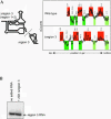
(A) Folding of a truncated PSIV IRES RNA. On the left is a cartoon of the construction of two truncated PSIV IRES mutants: Δregion 3 (nucleotides 6002–6149) and region 3 (nucleotides 6149–6195). Comparison of the hydroxyl radical probing profile of full-length PSIV IRES and Δregion 3 is shown on the right. The analysis and color scheme is identical to that in Figure 2 ▶. An asterisk denotes the location of an apparent difference in the cleavage pattern, but this change was not observed on all gels. (B) Native gel of labeled region 3 RNA with and without 20-fold molar excess of Δregion 3. The lack of a shift upon addition of the larger RNA indicates no in trans binding.
FIGURE 6.
FIGURE 6.
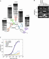
Analysis of the folding and 40S subunit binding of mutant IGR IRES RNAs. (A) Diagram of mutants used to link the IRES fold with 40S subunit binding. For each of the pseudoknots, we altered one side of the base-pairing interaction to its Watson–Crick complement. For the stem–loops, we substituted stable GAAA tetraloops, as these should maintain the integrity of the secondary structure. Adjacent to each of the five mutants is a portion of a hydroxyl radical probing gel of that mutant (bottom of the gel is ~nt 6084). All but mutants ΔPK III and ΔPK II show wild-type protection upon folding. (B) Native gel electrophoresis of wild-type PSIV IRES RNA and the five mutants shown in part A in the absence and in the presence of magnesium. (C) Binding curves of wild-type PSIV IRES RNA and the five mutants. Only ΔPK I binds as wild-type.

Similar articles

Cited by

References

    1. Adams, P.L., Stahley, M.R., Kosek, A.B., Wang, J., and Strobel, S.A. 2004. Crystal structure of a self-splicing group I intron with both exons. Nature 430: 45–50. - PubMed
    1. Bonnal, S., Boutonnet, C., Prado-Lourenco, L., and Vagner, S. 2003. IRESdb: the Internal Ribosome Entry Site database. Nucleic Acids Res. 31: 427–428. - PMC - PubMed
    1. Cate, J.H., Gooding, A.R., Podell, E., Zhou, K., Golden, B.L., Kundrot, C.E., Cech, T.R., and Doudna, J.A. 1996. Crystal structure of a group I ribozyme domain: Principles of RNA packing. Science 273: 1678–1685. - PubMed
    1. Celander, D.W. and Cech, T.R. 1991. Visualizing the higher order folding of a catalytic RNA molecule. Science 251: 401–407. - PubMed
    1. Christian, P.D. and Scotti, P.D. 1998. Picornaviruses of insects. In The insect viruses (eds. L.K. Miller and A. Ball), pp. 301–336. Plenum, New York.

LinkOut - more resources