. 2005 Feb 8;33(3):825-37.
doi: 10.1093/nar/gki185. Print 2005.

Splicing of human immunodeficiency virus RNA is position-dependent suggesting sequential removal of introns from the 5' end

Affiliations

Splicing of human immunodeficiency virus RNA is position-dependent suggesting sequential removal of introns from the 5' end

Jens Bohne et al. Nucleic Acids Res. .

Abstract

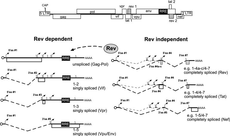
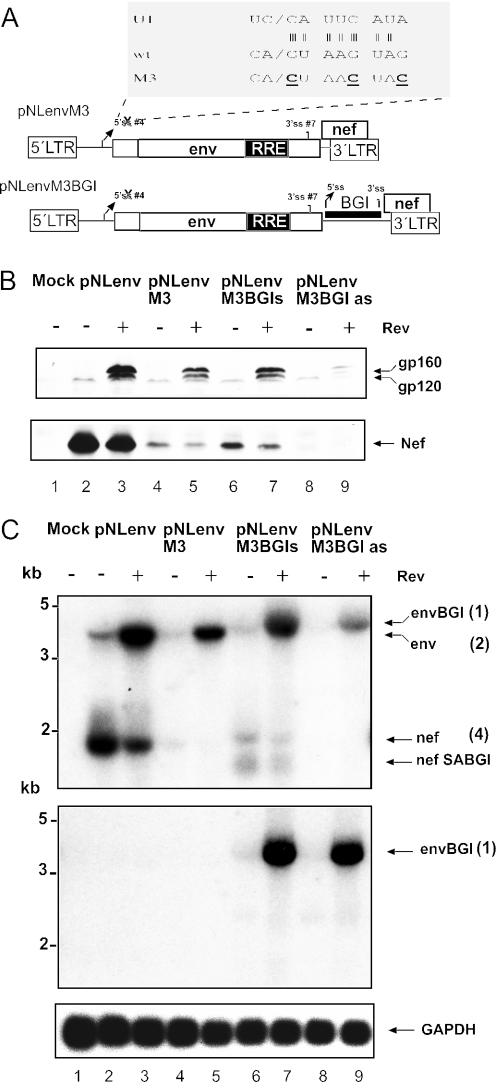
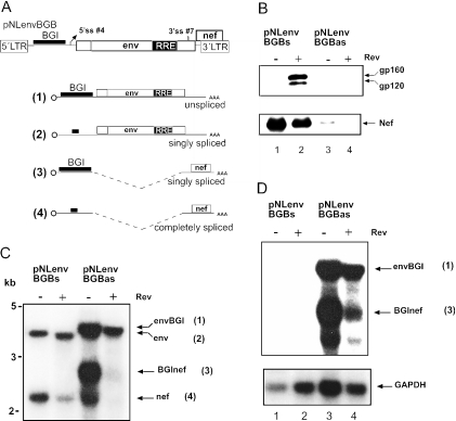
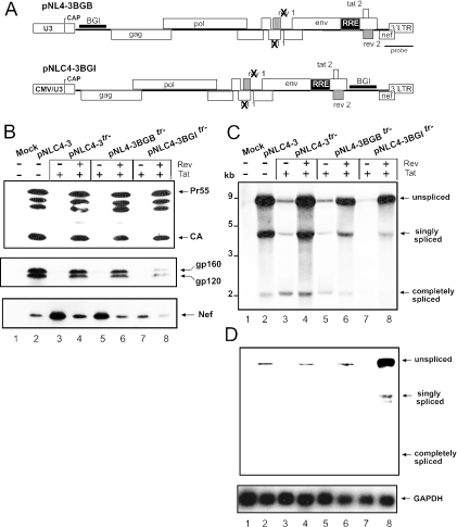
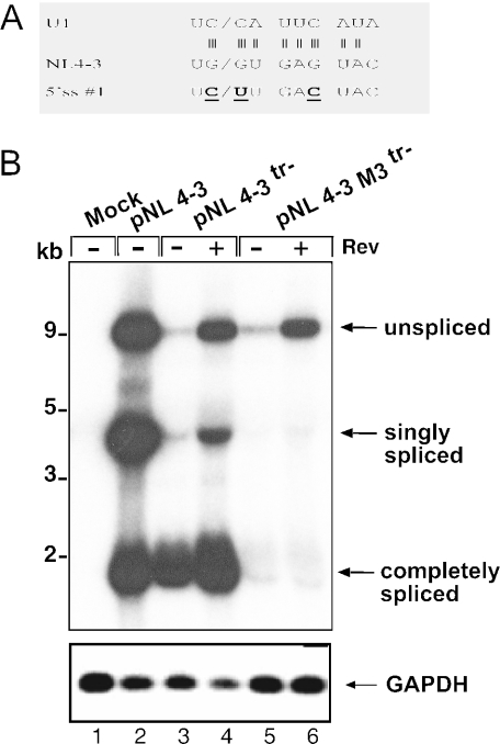
Transcription of the HIV-1 genome yields a single primary transcript, which is alternatively spliced to >30 mRNAs. Productive infection depends on inefficient and regulated splicing and appears to proceed in a tight 5' to 3' order. To analyse whether sequential splicing is mediated by the quality of splice sites or by the position of an intron, we inserted the efficient beta-globin intron (BGI) into the 3' region or 5'UTR of a subgenomic expression vector or an infectious proviral plasmid. RNA analysis revealed splicing of the 3' BGI only if all upstream introns were removed, while splicing of the same intron in the 5'UTR was efficient and independent of further splicing. Furthermore, mutation of the upstream splice signal in the subgenomic vector did not eliminate the inhibition of 3' splicing, although the BGI sequence was the only intron in this case. These results suggest that downstream splicing of HIV-1 RNAs is completely dependent on prior splicing of all upstream intron(s). This hypothesis was supported by the mutation of the major 5' splice site in the HIV-1 genome, which completely abolished all splicing. It appears likely that the tight order of splicing is important for HIV-1 replication, which requires the stable production of intron containing RNAs, while splicing of 3' introns on incompletely spliced RNAs would be likely to render them subject to nonsense-mediated decay.


Figures

Figure 1
Figure 1
Splicing pattern of HIV-1. The genomic organization of HIV-1 is depicted on top with open boxes representing ORFs and 5′ and 3′ LTRs, respectively. Classes of Rev-dependent RNAs are shown below on the left side, Rev-independent RNAs on the right side. A circle represents the 5′ cap, translated reading frames are depicted as open boxes, the RRE as a black box, and 5′ and 3′ss as arrows and vertical lines, respectively. The nomenclature of splice sites was adapted from Purcell and Martin, (8). Below each RNA, the splice sites used for its generation and the translation product are identified.
Figure 2
Figure 2
The subgenomic pNLenv expression system. (A) Schematic depiction of pNLenv (top). The vpu, env and nef ORFs as well as the 5′ and 3′ LTRs and the localization of the probe used for northern blot analysis are shown. The middle and bottom drawings depict the unspliced (middle) and spliced RNA produced from pNLenv. (B) Indirect immunofluorescence analysis of HeLa P4 cells transfected with pNLenv with (top) or without (bottom) a Rev expression plasmid. A Tat expression plasmid was cotransfected to induce LTR-dependent transcription. Cells were stained with antiserum against Env (top) or Nef (bottom). (C) Western blot analysis of transfected HeLa P4 cells. Cells were mock-transfected (lane 3) or transfected with pNLenv and a Tat expression plasmid either with (lane 2) or without (lane 1) a Rev expression plasmid. Blots were stained with antiserum against Env (top) or Nef (bottom). The viral glycoprotein precursor gp160 and the surface glycoprotein gp120 (which contains the epitope for the antibody) as well as the Nef protein are identified on the right. (D) Northern blot analysis of RNA from cell extracts from the same transfection as in panel C. Ten μg of RNA was separated on a denaturing gel. The blot was hybridized with a 3′ LTR-specific probe as indicated in panel A. Re-hybridization with a GAPDH-specific probe served as the loading control (bottom panel). The unspliced and spliced RNAs are identified on the right and molecular mass standards are shown on the left.
Figure 3
Figure 3
Splicing of a heterologous intron in the 3′ region of HIV-1. (A) Schematic depiction of the construct (top) and the RNA products. The second intron of the rabbit BGI was cloned in sense or antisense orientation between the reading frames of env and nef of pNLenv. Drawings (1–4) show the potential RNA species plus the shorter product (X). Identification of ORFs, cap and splice signals is as shown in Figure 4; the BGI sequence and the remaining heterologous sequence after splicing of this intron are shown as thick lines. Primers for RT–PCR analysis (see Figure 5) are also indicated in the top panel. (B) Western blot analysis of HeLa P4 cells transfected with the plasmids identified above each lane. A Tat expression plasmid was cotransfected in all cases. The blot was stained and labeled as described in Figure 2B. (C) Northern blot analysis of cell extracts from the same transfection as in panel B. RNA separation and hybridization was performed as described in Figure 2C. The respective RNA species and their numbering in panel A is shown on the right. The shortest RNA species labeled nefSABGI (X) corresponds to the product of splicing from the HIV-1 5′ss #4 to the 3′ss of the BGI sequence and therefore, yields an RNA shorter than authentic nef mRNA. The blot was re-hybridized with a GAPDH-specific probe (bottom).
Figure 4
Figure 4
Analysis of the MINX intron in the 3′ region of pNLenv. The synthetic MINX intron was cloned into the 3′ region of pNLenv in the same position as described for the BGI sequence (pNLenvMINX). (A) Western blot analysis of HeLaP4 cells transfected with the constructs indicated above each lane. A Tat expression plasmid was cotransfected in all cases. HIV-1 specific proteins were identified as described in Figure 2B. (B) RT–PCR analysis of RNA isolated from the same transfection as in panel A. RNA was reverse transcribed and PCR amplified with primers hybridizing to the 3′ end of the env ORF and to the 3′ LTR (indicated in Figure 3A), and PCR products were separated on an agarose gel and stained with ethidium bromide. Specific products are identified on the right, an asterisk marks the position of the spliced product where the BGI sequence had been removed and the env intron had been retained. Molecular mass standards are given on the left. A parallel RT–PCR reaction with actin-specific primers (bottom panel) served as the RNA control. Control reactions without RT enzyme were negative (data not shown).
Figure 5
Figure 5
Mutation of the HIV-1 5′ss in pNLenv does not relieve the 3′ splice inhibition. (A) Schematic depiction of plasmids pNLenvM3 and pNLenvM3BGI. The sequence at the HIV-1 5′ss #4 (wt) and the complementary sequence of the U1 RNA is expanded on top. The three mutations introduced into the 5′ss are depicted below in bold and underlined (M3). (B) Western blot analysis of HeLa P4 cells transfected with the constructs indicated above each lane. A Tat expression plasmid was cotransfected in all cases. HIV-1 proteins are identified on the right as in Figure 2B. (C) Northern blot analysis of 10 μg of RNA obtained from the same transfection as in panel B. The upper panel was probed with an LTR-specific probe, the middle panel with a BGI-specific probe, and the bottom panel with a GAPDH-specific probe. The observed RNA species are identified as in Figure 3C; numbering is according to the drawing in Figure 3A.
Figure 6
Figure 6
Splicing of a heterologous intron in the 5′ region of HIV-1. (A) Schematic depiction of pNLenvBGB (top). The BGI sequence was cloned into the BssHII site located upstream of 5′ss #1. ORFs, splice signals and cap are identified as described in Figure 1. The possible RNA products are shown below. The thick line identifies the BGI sequence or the residual sequence after splicing of this intron. (B) Western blot analysis of HeLa P4 cells transfected with the plasmids indicated above each lane. A Tat expression vector was cotransfected in all cases. HIV-1 specific proteins were detected and labelled as described in Figure 2B. (C) Northern blot analysis of 10 μg of nuclear RNA derived from the same transfection as in panel B. Cells were fractionated and nuclear RNA was separated and hybridized with a probe specific for the HIV-1 LTR as described in Figure 2C. The respective RNA species and their numbering in panel A is shown on the right. (D) Re-hybridization of the same blot as in panel B with a BGI-specific probe (upper panel) and with a GAPDH-specific probe (lower panel).
Figure 7
Figure 7
Analysis of the splicing of a heterologous intron in the complete HIV-1 genome. (A) Schematic depiction of the proviral constructs containing the BGI sequence in the 5′ region (pNL4–3BGBtr−; top) or in the 3′ LTR (pNLC4–3BGItr−, bottom). The BGI sequence was inserted into the same positions as described for pNLenv. It is noted that the proviral clones with mutations in the tat and rev genes were used and Tat and Rev expression plasmids were cotransfected as indicated. In the case of pNLC4–3BGItr−, a derivative of pNL4-3 containing a cytomegalovirus promoter instead of the U3 region of the 5′ LTR was used (pNLC4-3). This construct had been shown to produce an infectious RNA with a titer comparable to pNL4-3 (44). (B) Hela P4 cells were transfected with the plasmids indicated above each lane. Cell extracts were analysed by western blot using an antiserum against the HIV-1 Gag derived capsid protein (CA). This antiserum detects the Gag precursor Pr55, several cleavage intermediates and the completely cleaved CA protein. Antisera against Env and Nef were used in the middle and lower panels, respectively. Viral proteins are marked on the right as in Figure 2B. (C) Northern blot analysis of RNA isolated from the same transfections as in panel B. The blot was hybridized with a LTR-specific probe as indicated in panel A. (D) Re-hybridization of the same blot as in panel C with a BGI (upper panel) and a GAPDH-specific (lower panel) probe. The positions of unspliced, singly spliced and completely spliced RNAs are shown on the right.
Figure 8
Figure 8
Mutation of the 5′ss #1 of HIV-1 abolishes splicing. (A) Schematic depiction of the wild-type sequence at the 5′ss #1 of pNL4-3 (middle), the complementary sequence of U1 (top) and the M3 mutations (bold and underlined; bottom). Vertical lines indicate hydrogen bonds between the 5′ss and U1. (B) Northern blot analysis of RNA from HeLa P4 cells transfected with the constructs indicated above each lane and hybridized with a HIV-1 LTR-specific probe. pNL4-3tr− is a derivative of pNL4-3 with mutations in the tat and rev genes. A Tat expression vector was cotransfected in the experiments shown in lanes 3–6. Unspliced, singly spliced and completely spliced RNAs are identified on the right, molecular mass standards are shown on the left. The bottom panel shows re-hybridization of the same blot with a GAPDH-specific probe.

References

    1. Coffin J.M., Hughes S.H., Varmus H.E. In: Retroviruses. Coffin J.M., editor. Cold Spring Harbor, NY: 1997. - PubMed
    1. Frankel A.D., Young J.A. HIV-1: fifteen proteins and an RNA. Annu. Rev. Biochem. 1998;67:1–25. - PubMed
    1. Pollard V.W., Malim M.H. The HIV-1 Rev protein. Annu. Rev. Microbiol. 1998;52:491–532. - PubMed
    1. Wodrich H., Krausslich H.G. Nucleocytoplasmic RNA transport in retroviral replication. Results Probl. Cell Differ. 2001;34:197–217. - PubMed
    1. Chang D.D., Sharp P.A. Regulation by HIV Rev depends upon recognition of splice sites. Cell. 1989;59:789–795. - PubMed

Publication types