. 2005 Feb 8;33(3):893-902.
doi: 10.1093/nar/gki234. Print 2005.

The p80 homology region of TEP1 is sufficient for its association with the telomerase and vault RNAs, and the vault particle

Affiliations

The p80 homology region of TEP1 is sufficient for its association with the telomerase and vault RNAs, and the vault particle

Michael J Poderycki et al. Nucleic Acids Res. .

Abstract

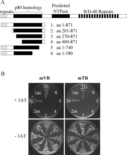
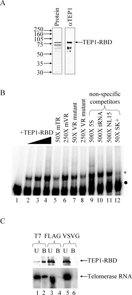
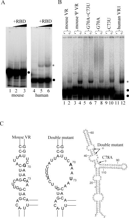
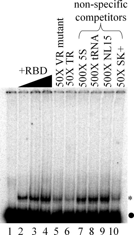
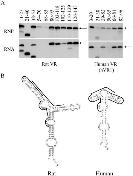
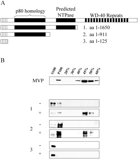
TEP1 is a protein component of two ribonucleoprotein complexes: vaults and telomerase. The vault-associated small RNA, termed vault RNA (VR), is dependent upon TEP1 for its stable association with vaults, while the association of telomerase RNA with the telomerase complex is independent of TEP1. Both of these small RNAs have been shown to interact with amino acids 1-871 of TEP1 in an indirect yeast three-hybrid assay. To understand the determinants of TEP1-RNA binding, we generated a series of TEP1 deletions and show by yeast three-hybrid assay that the entire Tetrahymena p80 homology region of TEP1 is required for its interaction with both telomerase and VRs. This region is also sufficient to target the protein to the vault particle. Electrophoretic mobility shift assays using the recombinant TEP1 RNA-binding domain (TEP1-RBD) demonstrate that it binds RNA directly, and that telomerase and VRs compete for binding. VR binds weakly to TEP1-RBD in vitro, but mutation of VR sequences predicted to disrupt helices near its central loop enhances binding. Antisense oligonucleotide-directed RNase H digestion of endogenous VR indicates that this region is largely single stranded, suggesting that TEP1 may require access to the VR central loop for efficient binding.


Figures

Figure 1
Figure 1
Yeast three-hybrid analysis of TEP1 deletions and vault/telomerase RNAs. (A) TEP1 truncations were made using amino acids 1–871 as a starting point, since this region of human TEP1 interacts with human vault and telomerase RNAs in the yeast three-hybrid system (16). (B) Yeast strain L40i was transformed with plasmids expressing the above TEP1 deletions and murine vault or telomerase RNA in either sense (s) or antisense (as) orientations. Yeast were grown on synthetic drop-out plates lacking uracil, leucine and histidine, and containing 5 mM 3-aminotriazole (+3AT) or synthetic media lacking only uracil and leucine (−3AT). Growth on +3AT media indicates a TEP1–RNA interaction. No TEP1 deletions interacted with antisense RNA constructs (data not shown), but this is shown above only for those TEP1 deletions that interact with RNA in the sense orientation (constructs 1 and 2).
Figure 2
Figure 2
Partially purified murine TEP1–RBD interacts with TR in vitro but has limited specificity. (A) Amino acids 201–871 of murine TEP1 were expressed in E.coli and partially purified using the hexahistidine tag derived from the pET28a vector. Shown are a Coomassie-stained gel (left panel) and western blot (right panel) using anti-TEP1 polyclonal antibodies. (B) EMSA of nt 1–223 of mouse TR incubated with TEP1–RBD. Five fmol 32P-labeled probe was incubated with 0, 5, 20 and 80 ng TEP1–RBD (lanes 1–4). Probe was competed off with 50× full-length TR (lane 5) and increasing amounts of mVR double point mutant (lanes 7 and 8), but not wild-type mVR (lane 6). The 250× unlabeled 5S Ribosomal RNA, tRNAphe or an artificial RNA (NL15) do not compete with the probe (lanes 7–9), but 50× excess of an artificial RNA derived from the pBluescript polylinker region does compete efficiently (lane 10). Labeled RNA is indicated with a black dot and shifted complexes are indicated with an asterisk. (C) Immunoprecipitation of TEP1–RBD from binding reactions using anti-T7 monoclonal antibody co-immunoprecipitates the TR transcript. Equivalent amounts of the bound (B) and unbound (U) fraction were analyzed by either western blot using anti-TEP1 polyclonal antibody (upper panel) or by fractionation on a 10% acrylamide/8 M urea gel (lower panel). The latter gel was dried and radioactive bands visualized by phosphorimager analysis. Antibodies to the T7 epitope (lanes 1 and 2), but not antibodies to the FLAG (lanes 3 and 4) and VSVG (lanes 5 and 6) epitopes, immunoprecipitated both TEP1–RBD and TR.
Figure 3
Figure 3
Mutagenesis of murine VR enhances its binding to TEP1–RBD. (A) EMSA of VR–TEP1–RBD complexes. Binding reactions contained 5 fmol of 32P-labeled murine VR (left panel) or human VR1 (right panel) incubated with 0 (lanes 1 and 4), 20 (lanes 2 and 5) and 80 (lanes 3 and 6) ng of TEP1–RBD. (B) EMSA of VR probes (5 fmol) without (−) or with (+) 80 ng TEP1–RBD in the binding reaction. Wild-type mouse VR interacts poorly or not at all with TEP1–RBD (lanes 1 and 2), but a highly related sequence in the mouse genome that is not expressed binds more strongly (lanes 3 and 4). A VR double point mutant G70A, C73U (lanes 5 and 6) binds more strongly to TEP1–RBD. Also shown are the corresponding single mutants in mouse VR (lanes 7–10) and human VR1 (lanes 11 and 12). Labeled RNAs are indicated with a black dot and shifted complexes are indicated with an asterisk. (C) The thermodynamically predicted secondary structure of the wild-type (left structure) and double point mutant (center structure) mouse VR, focusing on the regions of interest only. Also shown is the entire predicted structure of mouse VR, along with the position of the double point mutant and the C78 to A mutation, which has enhanced binding.
Figure 4
Figure 4
The mVR point mutant competes with TR for binding to TEP1–RBD. EMSA of mVR double point mutant incubated with 0, 5, 20 and 80 ng of TEP1–RBD (lanes 1–4). Unlabeled mVR mutant or mouse TR (50×) competes with the labeled probe (lanes 5 and 6). Three irrelevant RNAs do not compete with TR for binding to TEP1–RBD (lanes 7–9), but the pBluescript transcript does compete efficiently (lane 10) as in Figure 2. Labeled RNA is indicated with a black dot and shifted complexes are indicated with an asterisk.
Figure 5
Figure 5
RNase H digests of vault-bound and deproteinated VR. (A) Oligodeoxynucleotide-directed RNase H cleavage of the VR. P100 extract (RNP) or deproteinized P100 (RNA) purified from rat fibroblasts (left panel) or HeLa cells (right panel) was incubated with the indicated antisense ODN and RNase H. RNA was extracted from each sample and fractionated on a 10% denaturing polyacrylamide gel. The gel was electroblotted to a Zeta GT+ membrane and probed with the randomly primed VR gene. Intact VR is indicated by the arrow, and lower bands represent cleaved VR. (B) Thermodynamic prediction of rat VR and human VR1 secondary structures. The lines indicate regions of single-stranded RNA based upon RNase H digests of vault-associated VR.
Figure 6
Figure 6
The p80 homology region of TEP1 contains the vault interaction domain. (A) TEP1 truncations were co-expressed with the MVP in Sf9 insect cells using a baculovirus expression vector. (B) MVP forms vaults when expressed in insect cells, which are found entirely in the high-speed pellet (P100) of cell lysates and can be purified to near-homogeneity. Vault-interacting proteins will co-purify with vaults. Vaults were further enriched, and as a final step, samples were loaded onto a 20–60% sucrose gradient and centrifuged for 16 h. Sucrose fractions were pelleted, resuspended in 20 mM MES buffer, pH 6.5 and analyzed by western blot. Vaults fractionate largely in the 45% sucrose layer (upper panel). In the absence (−) of MVP-baculovirus co-infection, TEP1 truncations fractionate in either the high-speed supernatant (S100), P100, or both, and are lost in the purification scheme before the sucrose gradient step (1, 2, and 3, upper panels). When MVP is present (+), TEP1 truncations able to interact with vaults are found largely in the 45% sucrose layer (column 1, 2, and 3, lower panels).

References

    1. Kedersha N.L., Rome L.H. Isolation and characterization of a novel ribonucleoprotein particle: large structures contain a single species of small RNA. J. Cell. Biol. 1986;103:699–709. - PMC - PubMed
    1. Kedersha N.L., Miquel M.C., Bittner D., Rome L.H. Vaults. II. Ribonucleoprotein structures are highly conserved among higher and lower eukaryotes. J. Cell. Biol. 1990;110:895–901. - PMC - PubMed
    1. Kong L.B., Siva A.C., Rome L.H., Stewart P.L. Structure of the vault, a ubiquitous cellular component. Structure Fold. Des. 1999;7:371–379. - PubMed
    1. Stephen A.G., Raval-Fernandes S., Huynh T., Torres M., Kickhoefer V.A., Rome L.H. Assembly of vault-like particles in insect cells expressing only the major vault protein. J. Biol. Chem. 2001;276:23217–23220. - PubMed
    1. Chugani D.C., Rome L.H., Kedersha N.L. Evidence that vault ribonucleoprotein particles localize to the nuclear pore complex. J. Cell. Sci. 1993;106:23–29. - PubMed

Publication types

MeSH terms