

Review
doi: 10.1128/EC.4.2.225-229.2005.
Polarisome meets spitzenkörper: microscopy, genetics, and genomics converge
Affiliations
- PMID: 15701784
- PMCID: PMC549335
- DOI: 10.1128/EC.4.2.225-229.2005
Item in Clipboard
Review
Polarisome meets spitzenkörper: microscopy, genetics, and genomics converge
Eukaryot Cell.
2005 Feb.
No abstract available
Figures
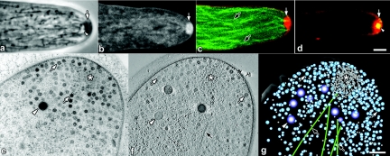
(a to d) Live-cell imaging of growing hyphal tips of N. crassa (a to c) and A. nidulans (d). Scale bars = 2.5 μm. (a) Digitally enhanced phase-contrast image of an unstained hypha showing Spk composed of a phase-dark cloud of secretory vesicles (arrow) and a phase-bright core (arrowhead) (M. Uchida and R. W. Roberson, unpublished data). (b) Confocal image of a hypha stained with FM4-64 showing pronounced staining of vesicles (arrow) within the Spk (P. C. Hickey and N. D. Read, unpublished data). (c) Confocal image colabeled with β-tubulin-GFP (green, black arrow) and FM4-64 (red, white arrow) showing microtubules extending into Spk. (d) Confocal image colabeled with SepA-GFP and FM4-64 (red, arrow) showing colocalization of SepA (yellow, arrowhead). In the central region of Spk is the region occupied by microvesicles (see panels e to g) (E. R. Kalkman and N. D. Read, unpublished data). (e to g) TEM of a hyphal tip of A. nidulans. A cluster of microvesicles within the Spk core (asterisks) surrounded by apical vesicles (white arrows), Woronin bodies (white arrowheads), and microtubules (black arrows) are indicated in these images. (e) Standard transmission electron micrograph (60-nm-thick section). (f) Electron tomographic section (∼3-nm z-axis resolution) of a dual-axis tomogram taken at 200 kV. (g) Model of panel f showing the 3D distribution of apical cytoplasm through an ∼170-nm-thick section (Uchida and Roberson, unpublished). Scale bar = 250 nm.
References
-
- Agrios, G. N. 1997. Plant Pathology, 4th ed. Academic Press, London, United Kingdom.
-
- Bartnicki-García, S., and E. Lippman. 1969. Fungal morphogenesis: cell wall construction in Mucor rouxii. Science 165:302-304. - PubMed
-
- Bartnicki-Garcia, S., F. Hergert, and G. Gierz. 1989. Computer simulation of fungal morphogenesis and the mathematical basis for hyphal tip growth. Protoplasma 153:46-57.
-
- Bartnicki-García, S. 2002. Hyphal tip growth: outstanding questions, p. 29-58 In H. D. Osiewacz (ed.), Molecular biology of fungal development. Marcel Dekker, Inc., New York, N.Y.
-
- Bartnicki-Garcia, S., C. E. Bracker, E. Reyes, and J. Ruiz-Herrera. 1978. Isolation of chitosomes from taxonomically diverse fungi and synthesis of chitin microfibrils in vitro. Exp. Mycol. 2:173-192.
Publication types
MeSH terms
LinkOut - more resources
Full Text Sources
Medical
