. 2005 Jun;288(6):F1153-63.
doi: 10.1152/ajprenal.00008.2005. Epub 2005 Feb 8.

Mispolarization of desmosomal proteins and altered intercellular adhesion in autosomal dominant polycystic kidney disease

Affiliations

Mispolarization of desmosomal proteins and altered intercellular adhesion in autosomal dominant polycystic kidney disease

Melina Silberberg et al. Am J Physiol Renal Physiol. 2005 Jun.

Abstract

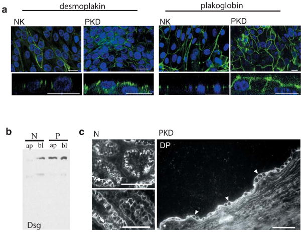
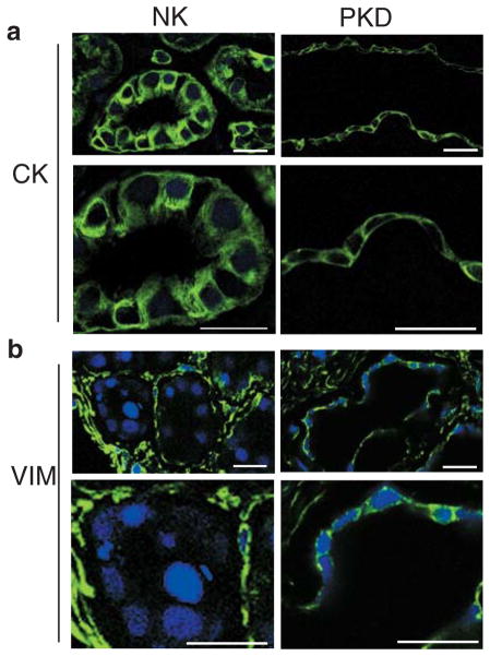
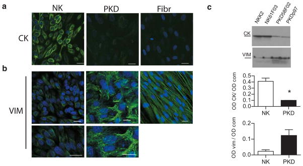
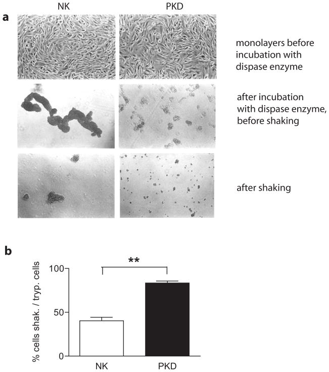
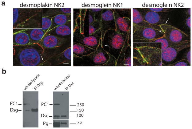
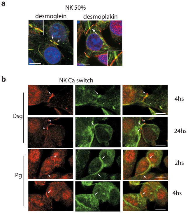
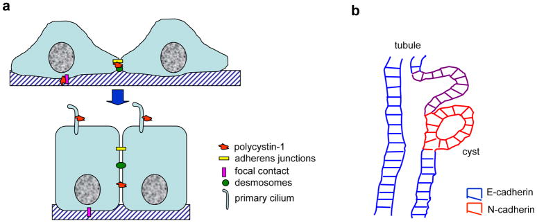
Polycystin-1, the product of the major gene mutated in autosomal dominant polycystic kidney disease (ADPKD), has been shown to associate with multiple epithelial cell junctions. Our hypothesis is that polycystin-1 is an important protein for the initial establishment of cell-cell junctions and maturation of the cell and that polycystin-1 localization is dependent on the degree of cell polarization. Using laser-scanning confocal microscopy and two models of cell polarization, polycystin-1 and desmosomes were found to colocalize during the initial establishment of cell-cell contact when junctions were forming. However, colocalization was lost in confluent monolayers. Parallel morphological and biochemical evaluations revealed a profound mispolarization of desmosomal components to both the apical and basolateral domains in primary ADPKD cells and tissue. Studies of the intermediate filament network associated with desmosomes showed that there is a decrease in cytokeratin levels and an abnormal expression of the mesenchymal protein vimentin in the disease. Moreover, we show for the first time that the structural alterations seen in adherens and desmosomal junctions have a functional impact, leaving the ADPKD cells with weakened cell-cell adhesion. In conclusion, in this paper we show that polycystin-1 transiently colocalizes with desmosomes and that desmosomal proteins are mislocalized as a consequence of polycystin-1 mutation.


Figures

Figure 1
Figure 1. Desmosomes are mispolarized in ADPKD cells in vitro and in situ
a. Confocal images of normal and ADPKD cells in culture labeled for desmosomal components (green). Upper panels: xy view to show desmosomal appearance at lateral membrane. Lower panels: xz view to show desmosomal distribution between apical and lateral cell domains. Desmoplakin labeling (two left panels) is seen only at the lateral domain of normal kidney cells (NK) but apical and lateral in ADPKD cells (PKD). Plakoglobin (two right panels) in normal (NK) and ADPKD cells (PKD) reveals same phenomenon. Nuclear staining, used as structural reference is seen in blue. Bars: 20 μm (except first panel: 5μm) b. Western blot of streptavidin-precipitated, biotinylated proteins, blotted with anti-desmoglein antibody. Only basolateral protein (bl) was recovered in normal kidney cells (N), while basolateral (bl) and apical (ap) desmoglein is seen in ADPKD cells (P). C. Confocal image of cryosections of normal (two left panels) and polycystic (right panel) tissue. Labeling against desmoplakin shows apical staining of the cyst lining epithelia in polycystic kidney tissue (arrowheads) while only lateral signal can be observed in normal tubules (arrows). Bars: 30 μm
Figure 2
Figure 2. Vimentin is anomalously expressed in cystic epithelia in situ
a. Cryosection of normal kidney showing cytokeratin (CK) staining in tubule cells (NK) Lower panel is a higher magnification of upper one. ADPKD kidney sectioned at a cyst level (PKD) shows that cells lining the wall cyst conserve cytokeratin expression (magnified in lower panel). Samples were imaged on Zeiss LSM510. Bars, 20 μm. b. Cryosection of normal tubules (NK) labeled against vimentin protein (VIM), show complete a absence of vimentin from their cytoplasm (magnified in lower panel). Kidney interstitial fibroblasts have a normal vimentin signal. Sections from a cyst of an ADPKD kidney (PKD) show the abnormal presence of vimentin in cyst-lining epithelial cells (detailed in lower panel). Samples were imaged on Zeiss LSM510. Bars, 20 μm.
Figure 3
Figure 3. ADPKD cells express an immature intermediate filament network even when fully confluent
a. Confocal images of primary normal kidney cells (NK) and polycystic kidney cells (PKD) in culture labeled against cytokeratin intermediate filament (CK). Normal kidney cells show normal cytokeratin labeling, while disease cells show very low signal. Confocal images of normal and polycystic kidney cells in culture, labeled against vimentin intermediate filament (VIM). Normal cells have very little vimentin signal (magnified in bottom panel), but polycystic cells, on the contrary, show a conspicuous vimentin filament network (amplified in lower panel). Normal human fibroblasts (Fibr) express exclusively vimentin. Bars, 20 μm. b. Immunoblots of lysates from two different samples each of normal (NK) and ADPKD (PKD) kidney cells probed for cytokeratin (CK) or vimentin (VIM). Below, quantification of cytokeratin and vimentin protein expression levels in normal and disease cells grown. The y-axis represents the intensity of the CK or the VIM band (OD CK) normalized according to the total protein loaded per lane as detected by Coomassie blue staining (OD com). CK graph: p=0.01. Vim graph: p=0.057.
Figure 4
Figure 4. Desmosomal mediated cell-cell adhesion is compromised in ADPKD cells
NK and PKD cell monolayers were incubated with dispase, released from the substrate and briefly shaken for a fixed time. a. Cells were imaged with a Zeiss, inverted light microscope. Top panels show NK and PKD cells forming confluent monolayers (10X objective). Middle panels show normal kidney cells in large, interconnected sheets (NK cells), while ADPKD cells are in small aggregates (PKD) after incubation with dispase enzyme, but before shaking. Bottom panels show the same samples after shaking, where the difference is further exacerbated. b. Quantification of cells released after shaking was performed using a Beckman Coulter counter. Plotted are the percentages of single or doublet cells after shaking as a function of the total number of cells assayed by trypsinization. 83.4 ± 2.20 % of PKD cells (black) were present as single or doublet cells after shaking, while only 40.3 ± 3.95 % of NK cells (white) were present as single or doublet cells after the same procedure. P= 0.005.
Figure 5
Figure 5. Polycystin-1 and desmosomal proteins are segregated in fully confluent normal kidney cells
a. Confocal images of primary normal human kidney cells grown on filters at 100% confluence for three days co-labeled for polycystin-1 (red) and desmosomal components (green). No colocalization between polycystin-1 and desmoplakin (left panel) was seen. Desmoglein staining of normal kidney cells (NK1 and NK2) from two different patients shows no overlap with polycystin-1 signal (arrows). Occasional colocalization could be seen in membrane regions where desmosome and polycystin-1 staining gave a homogeneous pattern (arrowhead, right panel). Insets show a magnification of the regions denoted by arrows. Each image depicts a single confocal section acquired at the level of the nucleus. Bars, 5 μm. b. Normal kidney cells were lysed and subjected to immunoprecipitation for different desmosomal proteins. Subsequent western blotting was used to probe for the presence of polycystin-1. Polycystin-1 failed to be recovered in immunoprecipitates of desmocollin or desmoglein. A blot for plakoglobin was included as a positive control for the stability of desmosomal protein-protein interactions to the lysis procedure.
Figure 6
Figure 6. Polycystin-1 and desmosomal proteins transiently colocalize when normal kidney cells are at 50% confluence
Confocal images of normal primary human kidney cells taken at early stage of cell-cell contact. Co-labeling of polycystin-1 (red) and desmosomal components (green) was carried out. a. Cells processed at 50% confluence show an overlapping pattern (arrows) between polycystin-1 and desmoglein (left panel) or desmoplakin (right panel). Insets show a magnification of membrane region marked with arrows. Bars, 10 μm. b. Cells were subjected to a calcium switch assay and processed at different time points after the calcium switch. Desmoglein staining (upper two rows) shows clear colocalization with polycystin-1 at 4 h following the calcium switch. This colocalization is lost after 24 h. Plakoglobin labeling (lower two rows) shows colocalization with polycystin-1 at 2 and 4 h after the switch to normal calcium. Bars, 10 μm.
Figure 7
Figure 7. Models
a. Schematic representation of our hypothesis of changing polycystin-1 distributions with cell polarization. In subconfluent, polarizing monolayers polycystin-1 is expressed at the basal and lateral membranes to facilitate the establishment of focal adhesions, as well as adherens junctions and desmosomes, possibly by mediating localized, regulated influx of calcium. Upon reaching full confluence and establishment of fully mature adhesive junctions, polycystin-1 no longer interacts with those junctions at the basolateral membranes and is relocated to the primary cilium where it is important in mechanosensory signal transduction and may participate in the maintenance of cell polarity. b. Schematic representation of how altered cell-cell adhesion may contribute to cystogenesis. Cells suffering a second site mutation in polycystin-1 or –2 begin to proliferate, but fail to reestablish a fully polarized epithelium. Weakened cell-cell adhesion caused by improper desmosome assembly coupled with the increased migratory potential of N-cadherin expressing cystic epithelia likely are envisaged to contribute to the extrusion of the cystic cells into the underlying parenchyma. The closure of the cyst may be driven by the normal tubular epithelia attempting to reconnect with their normal counterparts to reestablish a contiguous monolayer.

Similar articles

Cited by

References

    1. Arnaout MA. Molecular genetics and pathogenesis of autosomal dominant polycystic kidney disease. Annu Rev Med. 2001;52:93–123. - PubMed
    1. Sutters M, Germino GG. Autosomal dominant polycystic kidney disease: molecular genetics and pathophysiology. J Lab Clin Med. 2003;141:91–101. - PubMed
    1. Grantham JJ, Geiser JL, Evan AP. Cyst formation and growth in autosomal dominant polycystic kidney disease. Kidney Int. 1987;31:1145–1152. - PubMed
    1. Carone FA, Makino H, Kanwar YS. Basement membrane antigens in renal polycystic disease. Am J Pathol. 1988;130:466–471. - PMC - PubMed
    1. Wilson PD. Polycystin: new aspects of structure, function, and regulation. J Am Soc Nephrol. 2001;12:834–845. - PubMed

Publication types

MeSH terms

LinkOut - more resources