Comparative Study
. 2005 May;16(5):2382-94.
doi: 10.1091/mbc.e04-10-0857. Epub 2005 Feb 9.

Vertebrate Nup53 interacts with the nuclear lamina and is required for the assembly of a Nup93-containing complex

Affiliations
Comparative Study

Vertebrate Nup53 interacts with the nuclear lamina and is required for the assembly of a Nup93-containing complex

Lisa A Hawryluk-Gara et al. Mol Biol Cell. 2005 May.

Abstract

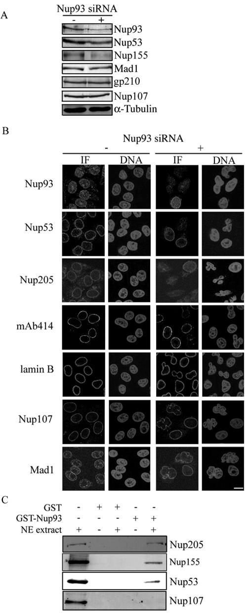
The nuclear pore complex (NPC) is an evolutionarily conserved structure that mediates exchange of macromolecules across the nuclear envelope (NE). It is comprised of approximately 30 proteins termed nucleoporins that are each present in multiple copies. We have investigated the function of the human nucleoporin Nup53, the ortholog of Saccharomyces cerevisiae Nup53p. Both cell fractionation and in vitro binding data suggest that Nup53 is tightly associated with the NE membrane and the lamina where it interacts with lamin B. We have also shown that Nup53 is capable of physically interacting with a group of nucleoporins including Nup93, Nup155, and Nup205. Consistent with this observation, depletion of Nup53 using small interfering RNAs causes a decrease in the cellular levels of these nucleoporins as well as the spindle checkpoint protein Mad1, likely due to destabilization of Nup53-containing complexes. The cellular depletion of this group of nucleoporins, induced by depleting either Nup53 or Nup93, severely alters nuclear morphology producing phenotypes similar to that previously observed in cells depleted of lamin A and Mad1. On basis of these data, we propose a model in which Nup53 is positioned near the pore membrane and the lamina where it anchors an NPC subcomplex containing Nup93, Nup155, and Nup205.


Figures

Figure 1.
Figure 1.
Localization of endogenous Nup53 to the NPC. (A) HeLa cells were processed for immunofluorescence microscopy and probed with affinity-purified rabbit antibodies directed against a putative vertebrate counterpart of Nup53 and a mouse mAb (mAb414) that binds a subset of FG-nups. Binding was detected with Cy3-and FITC-labeled secondary antibodies, respectively. Images of both fluorophores were acquired using a confocal microscope. Merged images are shown on the right. (B) HeLa cells were transfected with a plasmid encoding GFP-Nup53 and a stable cell line was isolated. Cells were fixed and probed with mAb414 as in A but detected with Cy3-labeled secondary antibodies. The cellular distribution of GFP-Nup53 (green), the mAb414 reactive nups (red), and a merged image are shown. Bar, 10 μm. (C) Proteins derived from indicated rat liver subcellular fractions were separated by SDS-PAGE and probed with Nup53-specific antibodies, as well as antibodies directed against the NPC proteins gp210 and Nup155. Nup53 was detected in fractions enriched in nuclei and nuclear envelopes.
Figure 2.
Figure 2.
Nup53 interacts tightly with the nuclear envelope membrane and lamina. (A) Purified rat liver NEs were extracted with 1 M NaCl, 2 M NaCl, 2 M urea, or 7 M urea and then centrifuged to produce a supernatant fraction (S) and a membrane pellet (P) fraction. After separation of proteins by SDS-PAGE, fractions were analyzed by Western blotting using antibodies that bind the indicated proteins. (B) Alternatively, NE membranes were extracted with Triton X-100, and an insoluble NPC and lamina-containing fraction was recovered by centrifugation. This extracted NE (eNE) fraction was then treated with increasing concentrations of NaCl to release nups from the lamina. After centrifugation to produce supernatant and pellet fractions, proteins were analyzed by Western blotting as in A. (C) A previously described extraction protocol of NEs (Matunis et al., 1996; Cronshaw et al., 2002) that concludes with empigen BB-induced release of NPCs from the lamina was also used to evaluate the association of Nup53 with the lamina. After extraction with empigen BB and centrifugation, proteins in a nup-enriched (S) fraction and a lamin-enriched (P) fraction were analysis by Western blotting using the antibodies specific to the indicated proteins.
Figure 3.
Figure 3.
Identification of Nup53-interacting partners using in vitro binding assays. (A) Purified rat liver NEs were extracted with 400 mM NaCl and 1% Triton X-100. After centrifugation to remove insoluble material, the supernatant fraction, containing nups and lamins, was diluted 3.75-fold (NE extract) and incubated with purified recombinant GST-Nup53 or GST alone immobilized on glutathione Sepharose 4B beads. After binding and washing steps, the bead-bound proteins were eluted, separated by SDS-PAGE, and detected with silver staining (SS). Several representative protein species that specifically bound Nup53 are indicated with an arrow. Samples were also analyzed by Western blotting (WB) using polyclonal antibodies directed against the indicated proteins or mAb414 to detect Nup358, Nup214, Nup153, and p62. (B) Purified recombinant Nup93 was incubated with GST-Nup53 or GST alone immobilized on glutathione Sepharose 4B beads. After washing, beads were eluted with sample buffer. Proteins were separated by SDS-PAGE and analyzed by Coomassie Blue staining (CB) or Western blotting (WB) using antibodies directed against Nup93. The positions of mass markers in kilodaltons are indicated.
Figure 4.
Figure 4.
In vivo depletion of Nup53 using small interfering RNAs. (A) HeLa cells were incubated for 48 h in the presence of either transfection reagent alone (-) or siRNA (+) corresponding to nonsense siRNAs or Nup53 siRNAs. Samples were then collected and the cellular levels of Nup53 and α-tubulin (for comparison of total protein loaded in each fraction) were analyzed by Western blotting of total cell extracts. (B) HeLa cells growing on coverslips were incubated for 48 h with transfection reagent alone (mock), nonsense siRNAs (nonsense), or Nup53 (Nup53) siRNAs and processed for immunofluorescence. Cells were probed with anti-Nup53 antibodies, and binding was detected with Cy3-labeled secondary antibodies. Nuclear DNA was detected by staining with the DNA-binding dye Hoechst. Bar, 10 μm.
Figure 5.
Figure 5.
HeLa cells depleted of Nup53 exhibit a growth delay. (A) Equal numbers of HeLa cells were transfected with either nonsense or Nup53-specific siRNA duplexes and incubated for the indicated times. At each time point, cells were collected, their viability was assessed using trypan blue and the number of viable cells was plotted versus time. Note, both nonsense and Nup53-specific siRNA treated cells showed a similar viability (>90%). The results shown are representative of three separate trials. (B) As in A, HeLa cells were transfected with either nonsense or Nup53-specific siRNA duplexes and incubated for the indicated times. Cells were then collected and processed for FACS analysis. The positions of the 2C and 4C peaks are indicated.
Figure 6.
Figure 6.
In vivo depletion of Nup53 leads to codepletion of a subset of interacting nups and the mitotic checkpoint protein Mad1. (A) Forty-eight hours after the transfection of HeLa cells with either nonsense (-) or Nup53 (+) siRNAs, cells were processed for indirect immunofluorescence (IF). Samples were probed with antibodies directed against the indicated proteins, and binding was detected with Cy3-labeled secondary antibodies. Mab414 binding was detected with FITC-labeled secondary antibodies. Nuclear DNA was detected by staining with the DNA-binding dye Hoechst (DNA). Bar, 10 μm. (B) Cells were transfected as described in A, total cell extracts were isolated, and proteins were separated by SDS-PAGE. Western blot analysis was performed using antibodies directed against the indicated proteins. Note, mAb414 was used to visualize the levels of p62, Nup153, and Nup214. Antibodies directed against α-tubulin were used for comparing total protein loads in each fraction.
Figure 7.
Figure 7.
In vivo depletion of Nup93 and the identification Nup93-interacting partners using in vitro binding assays. (A) Forty-eight hours after the transfection of HeLa cells with either nonsense (-)or Nup93 (+) siRNAs, total cell extracts were isolated, and proteins were separated by SDS-PAGE. Western blot analysis was performed using antibodies directed against the indicated proteins. Antibodies directed against α-tubulin were used for comparing total protein loads in each fraction. (B) Cells were transfected as described in A and processed for indirect immunofluorescence (IF). Samples were probed with antibodies directed against the indicated proteins, and binding was detected with Cy3-labeled secondary antibodies. Mab414 binding was detected with FITC-labeled secondary antibodies. Nuclear DNA was detected by staining with the DNA-binding dye Hoechst (DNA). Bar, 10 μm. (C) Purified recombinant GST-Nup93 or GST alone were immobilized on glutathione Sepharose 4B and incubated with or without rat liver NE extracts as described for Nup53 in Figure 3A. The bead-bound proteins were eluted, separated by SDS-PAGE, and analyzed by Western blotting using antibodies directed against the indicated nups.

Similar articles

Cited by

References

    1. Aitchison, J. D., Blobel, G., and Rout, M. P. (1995a). Nup120p: a yeast nucleoporin required for NPC distribution and mRNA transport. J. Cell Biol. 131, 1659-1675. - PMC - PubMed
    1. Aitchison, J. D., Rout, M. P., Marelli, M., Blobel, G., and Wozniak, R. W. (1995b). Two novel related yeast nucleoporins Nup170p and Nup157p: complementation with the vertebrate homologue Nup155p and functional interactions with the yeast nuclear pore-membrane protein Pom152p. J. Cell Biol. 131, 1133-1148. - PMC - PubMed
    1. Allen, N. P., Patel, S. S., Huang, L., Chalkley, R. J., Burlingame, A., Lutzmann, M., Hurt, E. C., and Rexach, M. (2002). Deciphering networks of protein interactions at the nuclear pore complex. Mol. Cell Proteomics 1, 930-946. - PubMed
    1. Belgareh, N., and Doye, V. (1997). Dynamics of nuclear pore distribution in nucleoporin mutant yeast cells. J. Cell Biol. 136, 747-759. - PMC - PubMed
    1. Belgareh, N. et al. (2001). An evolutionarily conserved NPC subcomplex, which redistributes in part to kinetochores in mammalian cells. J. Cell Biol. 154, 1147-1160. - PMC - PubMed

Publication types

MeSH terms