. 2005 Apr;16(4):1859-71.
doi: 10.1091/mbc.e04-08-0744. Epub 2005 Feb 9.

Nuclear translocation of integrin cytoplasmic domain-associated protein 1 stimulates cellular proliferation

Affiliations

Nuclear translocation of integrin cytoplasmic domain-associated protein 1 stimulates cellular proliferation

Henri-Noël Fournier et al. Mol Biol Cell. 2005 Apr.

Abstract

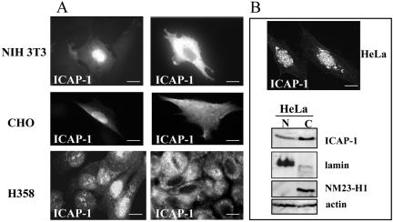
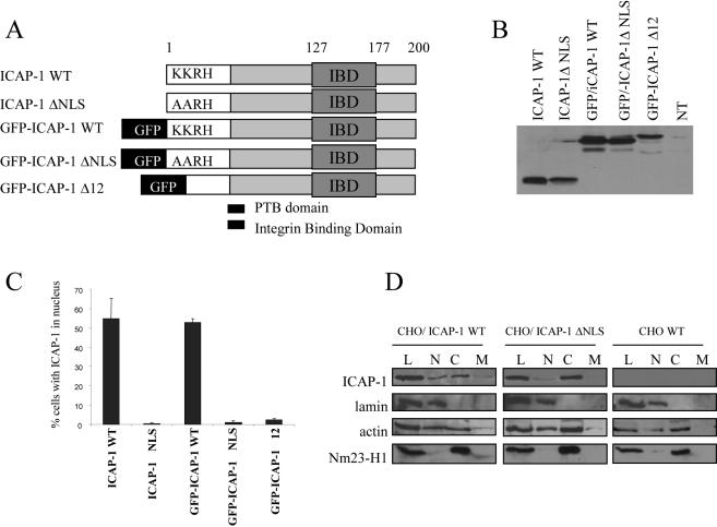
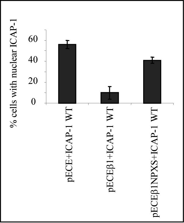
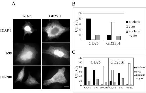
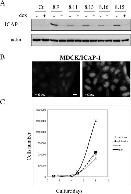
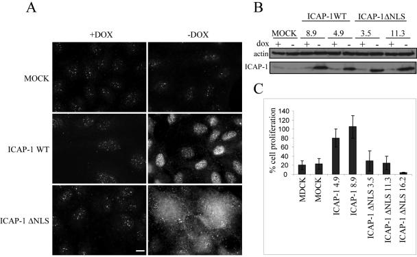
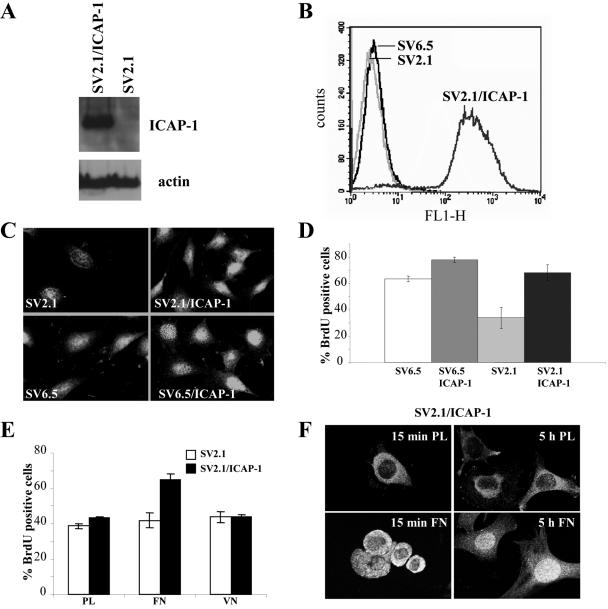
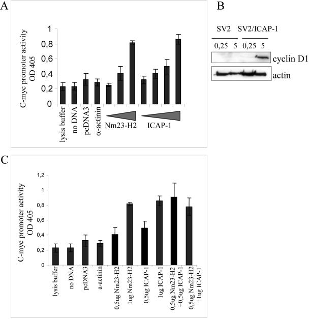
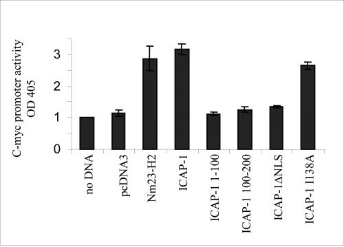
Integrin cytoplasmic domain-associated protein 1 (ICAP-1) has been shown to interact specifically with the beta1 integrin cytoplasmic domain and to control cell spreading on fibronectin. Interestingly, ICAP-1 also is observed in the nucleus, by immunocytochemical staining, and after biochemical cell fractionation, suggesting that it has additional roles that have yet to be determined. We show that the nucleocytoplasmic shuttling capability of ICAP-1 is dependent on a functional nuclear localization signal. In addition, overexpression of beta1 integrin strongly reduced this nuclear localization, suggesting that integrin activity could modulate ICAP-1 shuttling by sequestering it in the cytoplasm. Indeed, the nuclear localization of ICAP-1 is dependent on the stage of cell spreading on fibronectin, and we also show that ICAP-1 expression stimulates cellular proliferation in a fibronectin-dependent manner. This function is dependent on its nuclear localization. Moreover, ICAP-1 is able to activate the c-myc promoter in vitro. Together, these results demonstrate that ICAP-1 shuttles between the nucleus and cytoplasm in a beta1 integrin-dependent manner. It could act as a messenger that relays information from sites of integrin-dependent cell adhesion to the nucleus for controlling gene expression and cell proliferation.


Figures

Figure 1.
Figure 1.
Subcellular localization of ICAP-1. (A) Different types of cells were plated on coverslips coated with fibronectin. NIH 3T3 or CHO cells were transiently transfected by pcDNA3/ICAP-1. Twenty-four hours after transfection, the cells were fixed, permeabilized, and stained with anti-ICAP-1 polyclonal antibodies. The H358 cells stably transfected with pTREhyg/ICAP-1 were induced with 1 μg/ml doxycycline during 24 h before fixation, permeabilization, and immunostaining with anti-ICAP-1 polyclonal antibodies. Two photos per cell type are shown to visualize nuclear and cytoplasmic localization of ICAP-1. Bar, 10 μm. Endogenous ICAP-1 was visualized in HeLa cells by immunostaining with anti-ICAP-1 polyclonal antibodies (B) (bar, 10 μm.) and by Western blot (C) by using the fractionation procedure described under Materials and Methods. Fractionation and Western immunoblot analyses of HeLa cells confirmed the subcellular localization of ICAP-1 protein in the cytosol and nucleus. Fractions are labeled as follows: C, cytosol; N, nuclear. The various cell fractions were characterized by reprobing the blots for lamin as a nuclear marker, Nm23-H1 as a cytoplasmic marker and actin as a general marker.
Figure 2.
Figure 2.
Mutation in the NLS of full-length ICAP-1 results in the accumulation of the protein in the cytoplasm. (A) Schematic representation of the different mutants of ICAP-1 used for transfection. CHO cells were transfected with pcDNA3.1 plasmids that express different ICAP-1 proteins fused or not with GFP, with or without mutations in NLS sequence (ΔNLS corresponds to the change of the two lysine (KK6–7) of the NLS to alanine and delta 12 corresponds to the deletion of the 12 first amino acids of ICAP-1). (B) Proteins from cells 24 h after transfection were resolved by SDS-PAGE, and the wild-type and mutant ICAP-1 was revealed by Western blot by using ECL substrate. The NT track corresponds to a lysate of CHO cells transfected with the empty pcDNA3.1. (C) At 24 h posttransfection, the cells were prepared for immunofluorescence microscopy by using the anti ICAP-1 polyclonal antibodies, and ICAP-1 distribution was examined. Two patterns were visualized: either nuclear or diffuse. Scoring 100 cells for ICAP-1 protein distribution revealed that ICAP-1 WT resides in nucleus in 60% of cells, whereas mutation of NLS sequence results in the accumulation of ICAP-1 in the cytoplasm for almost 100% of the visualized cells. Experiments were independently reproduced at least three times. (D) Western blot analysis performed after cell fractionation of wild-type CHO and transfected CHO cells overexpressing either wild-type ICAP-1 (WT) or ICAP-1ΔNLS. Note the different distribution of ICAP-1WT and ICAP-1ΔNLS between the nuclear and the cytoplasmic fraction. Fractions are labeled as follows: C, cytoplasmic fraction; L, total lysate; M, membrane fraction; N, nuclear fraction.
Figure 3.
Figure 3.
β1 Integrin-dependent cytoplasmic translocation of ICAP-1. CHO cells were cotransfected with two different cDNAs, pcDNA3.1/ICAP and pECE/human β1 or pcDNA3.1/ICAP-1 and pECE or pcDNA3.1/ICAP-1 and pECE/human NPXS β1, as indicated in each histogram. The subcellular distribution of the ectopically expressed proteins was visualized by double immunostaining by using the anti-human β1 mAb 4B7R and polyclonal anti-ICAP antibodies. Two distribution patterns were visualized: either nuclear or diffuse. Scoring 100 cells for ICAP-1 protein distribution revealed that human β1 integrin cotransfection resulted in the sequestration of ICAP-1 in the cytoplasm for 90% of the visualized cells, whereas the cotransfection with NPXS human β1 integrin restores the dual cytoplasmic and nuclear distribution. Experiments were independently reproduced at least three times.
Figure 4.
Figure 4.
Subcellular localization of ICAP-1 is modulated by β1 integrin expression GD25 or GD25/β1 cells were transiently transfected with pcDNA3.1/ICAP-1, pcDNA3.1/ICAP-1(1-99), or pcDNA3.1/ICAP-1(100-200). (A) Subcellular distribution of the ectopically expressed proteins was visualized by immunofluorescence by using polyclonal anti-ICAP antibodies. Bar, 10 μm. (B and C) Three distribution patterns were visualized 24 h later as either nuclear, cytoplasmic, or both cytoplasmic and nuclear. Scoring 100 cells for ICAP-1 protein distribution revealed that cells devoid of β1 integrin sequestered ICAP-1 within nucleus (A and B). The 1–99 fragment bearing the NLS sequence was located in the nucleus (A and C) independently of the expression of β1 integrins, whereas the 100–200 fragment was sequestered in the cytoplasm (A and C).
Figure 5.
Figure 5.
Nuclear localization of ICAP-1 increases cell proliferation. (A) MDCK cells stably transfected with pTREhyg/ICAP-1 were induced for ICAP-1 expression by removing doxycycline during 48 h before lysis of the cells. Total proteins were subjected to gel electrophoresis and then transferred to PVDF membrane before processing of Western blot with the anti-ICAP-1 polyclonal antibodies. The same gel was blotted with anti-actin polyclonal antibodies for normalization. (B) Immunofluorescence of cells after induction shows a nuclear localization of ICAP-1. Bar, 10 μm. (C) After reducing the percentage of serum and maintaining cells in 4% serum, proliferation curves of MDCK cells containing pTREhyg/ICAP-1 (clone 8.9) or pTRE/hyg (ct) alone were registered in the absence (induction) or presence of 2 μg/ml doxycycline (+dox). The clone 8.9 expressing ICAP-1 shows an increase in cell proliferation after 5 d of induction.
Figure 6.
Figure 6.
Cellular distribution of ICAP-1 depends on its NLS sequence and exhibits differential effects on cell proliferation. (A) Subcellular localization of ICAP-1 was visualized by indirect immunofluorescence analysis with anti-ICAP-1 polyclonal antibodies in MDCK clones containing pTREhyg (MOCK cells) or pTREhyg/ICAP-1 (ICAP-1 WT) or pTREhyg/ICAP-1ΔNLS (ICAP-1ΔNLS) after induction (-dox) or without induction (+dox) with 2 μg/ml doxycycline. Bar, 10 μm. (B) After 7 d of induction (-dox), different clones expressing ICAP-1 WT (8.9 and 4.9) or ICAP-1ΔNLS (3.5 and 11.3) were lysed and expression of ICAP-1 with expected size was detected by Western blot with anti-ICAP-1 polyclonal antibodies. The same gel was blotted with anti-actin polyclonal antibodies for normalization. (C) The ratio of cell proliferation is shown for untransfected MDCK cells, MOCK cells, ICAP-1–overexpressing MDCK cells (clones 4.9 and 8.9), and ICAP-1ΔNLS–overexpressing MDCK cells (clones 3.5, 11.3, and 16.2) after 7 d of induction.
Figure 7.
Figure 7.
Loss of ICAP-1 decreases cell proliferation in osteoblasts. (A) ICAP-1–deficient osteoblasts (SV2.1) rescued or not with ICAP-1 were lysed with Laemmli buffer, and total cell lysates were resolved in SDS gel, transferred to a PVDF membrane, and probed with the monoclonal anti-ICAP-1 4D1D6 and anti-actin. (B) ICAP-1–deficient osteoblasts rescued with ICAP-1 (SV2.1/ICAP-1) were sorted by FACS by using EGFP fluorescence. (C) Immunostaining of ICAP-1 with polyclonal anti-ICAP-1 antibodies was performed in SV2.1, SV2.1/ICAP-1, SV6.5, and SV6.5/ICAP-1 cells. (D) BrdU incorporation was carried out in ICAP-1 null osteoblasts (SV2.1), ICAP-1-rescued SV2.1 cells (SV2.1/ICAP-1), wild-type osteoblasts (SV6.5), and SV6.5/ICAP-1. Cells that have incorporated BrdU were counted under the microscope by using anti-BrdU staining, and the percentage of labeled cells was estimated by 4,6-diamidino-2-phenylindole counterstaining. The results are the average of four experiments allowing us to estimate the SD. (D) BrdU incorporation was carried out in ICAP-1 null osteoblasts (SV2.1) or in ICAP-1-rescued SV2.1 cells (SV2.1/ICAP-1) plated either on polylysine (PL) or fibronectin (FN) or vitronectin (VN). Cells that have incorporated BrdU were counted under the microscope by using anti-BrdU staining. The results are the average of three experiments allowing us to estimate the SD. (E) SV2.1/ICAP-1 cells were allowed to spread onto FN or PL during 15 min (early stage of spreading) or 5 h (late stage of spreading), and then they were fixed, permeabilized, and stained with anti-ICAP-1 polyclonal antibodies. Visualization of a single section and image capture were done with a confocal microscope.
Figure 8.
Figure 8.
ICAP-1–mediated activation of the c-myc promoter. (A and C) CHO cells were transiently transfected with the c-myc-CAT P1 and P2 promoter construct (1 μg) along with increasing concentrations of Nm23-H2 expression plasmid (0, 0.5, and 1 μg) or ICAP-1 expression plasmid (0, 0.5, 0.7, and 1 μg). In all cases, the amount of DNA transfected was kept constant by the addition of the pcDNA3 plasmid (the vector backbone for Nm23-H2 or ICAP-1). Extracts were generated 48 h later and assayed for CAT expression. The results are the average of three experiments allowing us to estimate the SD. (B) SV2.1 and SV2.1/ICAP-1 cells were cultured for 24 h in 1% FCS before replating them onto fibronectin. After 15 min or 5 h of spreading, cells were washed with PBS and directly lysed onto petri dishes with RIPA buffer. An amount of 30 μg of proteins per lane was subjected to gel electrophoresis and then transferred to PVDF membrane before processing of Western blot with the anti-cyclin D1 mAb. The same gel was blotted with anti-actin polyclonal antibodies for normalization.
Figure 9.
Figure 9.
Activation of the c-myc promoter requires a functional NLS of ICAP-1. CHO cells were transiently transfected with the reporter construct pcmycCAT and the different constructs of ICAP-1. After 48 h, after lysis, CAT expression was determined according to the protocol of Roche Diagnostics.

Similar articles

Cited by

References

    1. Balda, M. S., and Matter, K. (2000). The tight junction protein ZO-1 and an interacting transcription factor regulate ErbB-2 expression. EMBO J. 19, 2024-2033. - PMC - PubMed
    1. Barberis, L., Wary, K. K., Fiucci, G., Liu, F., Hirsch, E., Brancaccio, M., Altruda, F., Tarone, G., and Giancotti, F. G. (2000). Distinct roles of the adaptor protein Shc and focal adhesion kinase in integrin signaling to ERK. J. Biol. Chem. 275, 36532-36540. - PubMed
    1. Barone, M. V., and Courtneidge, S. A. (1995). Myc but not Fos rescue of platelet-derived growth factor signalling block caused by kinase-inactive Src. Nature 378, 509-512. - PubMed
    1. Benaud, C. M., and Dickson, R. B. (2001). Regulation of the expression of c-Myc by β1 integrins in epithelial cells. Oncogene 20, 759-768. - PubMed
    1. Ben-Ze'ev, A., Shtutman, M., and Zhurinsky, J. (2000). The integration of cell adhesion with gene expression: the role of β-catenin. Exp. Cell Res. 261, 75-82. - PubMed

Publication types

MeSH terms

Substances