. 2005 Feb 22;102(8):3135-40.
doi: 10.1073/pnas.0500012102. Epub 2005 Feb 9.

Recruitment and interaction dynamics of plant penetration resistance components in a plasma membrane microdomain

Affiliations

Recruitment and interaction dynamics of plant penetration resistance components in a plasma membrane microdomain

Riyaz A Bhat et al. Proc Natl Acad Sci U S A. .

Abstract

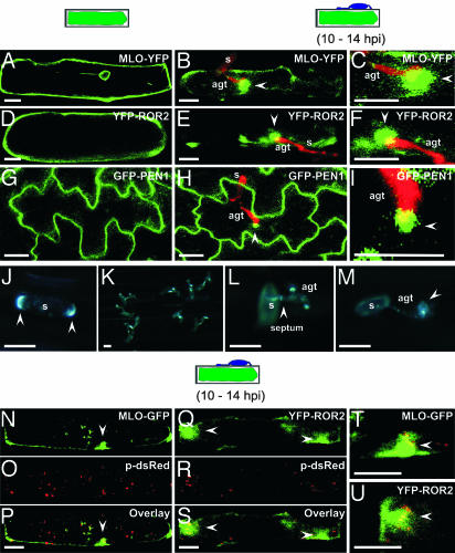
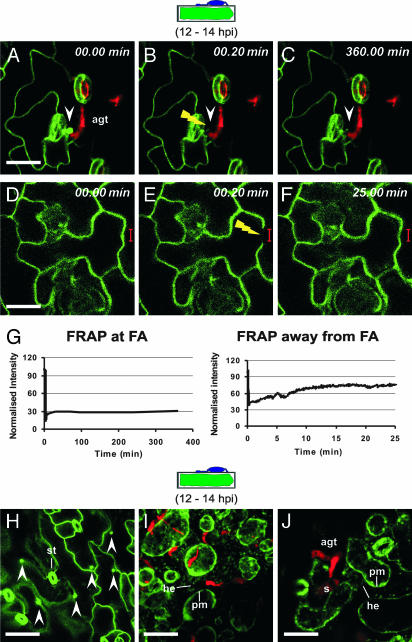
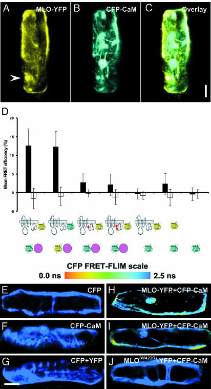
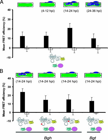
Many fungal pathogens must enter plant cells for successful colonization. Barley mildew resistance locus o (Mlo) is required for host cell invasion upon attack by the ascomycete powdery mildew fungus, Blumeria graminis f.sp. hordei, and encodes the founder of a family of heptahelical integral membrane proteins unique to plants. Recessively inherited loss-of-function mutant alleles (mlo) result in effective penetration resistance to all isolates of the biotrophic parasite. We used noninvasive fluorescence-based imaging to show that fluorescently tagged MLO protein becomes redistributed in the plasma membrane (PM) and accumulates beneath fungal appressoria coincident with the initiation of pathogen entry into host cells. Polarized MLO accumulation occurs once upon attack and appears to be independent of actin cytoskeleton function. Likewise, barley ROR2 syntaxin, a genetically defined component of penetration resistance to B. graminis f.sp. hordei, and a subset of predicted PM-resident proteins become redistributed to fungal entry sites. We previously identified calmodulin, a cytoplasmic calcium sensor, as an interactor and positive regulator of MLO activity and demonstrate here by FRET microscopy an increase in MLO/calmodulin FRET around penetration sites coincident with successful host cell entry. Our data provide evidence for the formation of a pathogen-triggered PM microdomain that is reminiscent of membrane microdomains (lipid rafts) induced upon attempted entry of pathogenic bacteria in animal cells.


Figures

Fig. 1.
Fig. 1.
Induced polarity in host cells and powdery mildew sporelings. (A–I) Pathogen-triggered cell polarity in plant cells. A barley epidermal cell expressing MLO-YFP (A–C) or YFP-ROR2 (D–F) in the absence and presence of Bgh sporelings (10–14 hpi). (G–I) Nonchallenged and Bgh-challenged Arabidopsis epidermal cells expressing GFP-PEN1. Epiphytic fungal structures (red) were stained with propidium iodide. Stages of fungal development on cross-sectioned epidermal cells are depicted schematically above the columns. Images shown are single focal planes. Close-up views of attempted fungal entry sites for MLO-YFP, YFP-ROR2, and GFP-PEN1 expressing cells are shown in C, F, and I, respectively. White arrowheads mark the positions of focally accumulated fusion proteins. s, conidiospore. (Scale bar, 20 μm.) (J–M) Filipin-mediated fluorescence in Bgh conidiospores and at pathogen entry sites. Bgh-challenged leaf sections were stained with filipin. A representative conidiospore before germination (J), overview of germinated conidia (K) and a close-up view of germinated conidiospores (L and M) are shown. In J and L, arrowheads mark opposite poles of the conidium and the septum between the spore body and AGT, respectively. Note the filipin-mediated fluorescence underneath the appressorium in the attacked host epidermal cell (M, marked by arrowhead). s, conidiospore. (Scale bar, 20 μm.) (N–U) A barley epidermal cell expressing either MLO-YFP or YFP-ROR2 together with HvADF3 S6A blocking actin cytoskeleton function. Disruption of actin cytoskeleton was monitored by coexpression of a peroxisome-targeted dsRed (p-dsRed) variant in the same cells. (N–P) Coexpression of MLO-GFP, p-dsRed, and HvADF3 S6A. (Q–S) Coexpression of YFP-ROR2, p-dsRed, and HvADF3. (T and U) Close-up views of attempted fungal entry sites in the same cells. The stage of fungal development on a cross-sectioned epidermal cell is depicted schematically above the columns. (Scale bar, 20 μm.)
Fig. 2.
Fig. 2.
Focal PEN1 accumulation is triggered once and occurs at the cytoplasmic face of biotic stress sites. (A–F) Arabidopsis leaves expressing GFP-PEN1 were inoculated with Bgh conidiospores. At 12 hpi, FRAP analysis was performed at (A–C) and away from (D–F) focal accumulation (FA) sites. White arrowheads mark the position of focally accumulated GFP-PEN1 and the yellow flash depicts the site of photobleaching. (G) Quantitative measure of fluorescence recovery over time at and away from FA sites. Note that the fluorescence recovery is incomplete (≈75%) away from FA sites because of continuous photobleaching during imaging. (H–J) Bgh-challenged Arabidopsis leaves stably expressing GFP-PEN1 were imaged at 12 hpi before (H) and after (I and J) plasmolysis. st, stomata; he, Hechtian threads. The stage of fungal development on a cross-sectioned epidermal cell is schematically depicted above the columns. (Scale bar, 20 μm.)
Fig. 3.
Fig. 3.
MLO interacts with CaM in nonchallenged barley epidermal cells. (A–C) Barley leaf epidermal cells were cobombarded with fluorescently tagged MLO (A) and CaM (B) and analyzed by confocal microscopy. The white arrowhead in A marks the position of focally accumulated MLO-YFP at an attempted fungal entry site. N, nucleus. Note that the images shown are 3D reconstructions from individual image stacks. (Scale bar, 20 μm.) (D) FRET-APB analysis of the interaction between fluorescently tagged MLO and CaM. Mean FRET efficiencies (black bars) and background FRET (white bars) ± SD from 50–100 sample sites are shown. MLO is depicted by its serpentine structure, CaM is a purple circle, and CFP and YFP fluorophores are cyan and yellow ribbon models, respectively. Red dots in the C-terminal tail of MLO symbolize the W423R and L420R/W423R amino acid substitutions in the CaMBD domain. (E–J) Pseudocolored fluorescence lifetime (FRET-FLIM) images of barley epidermal cells expressing combinations of CFP, YFP, and fluorescently tagged MLO and CaM forms. The bar at the top displays the scale of CFP donor fluorescence lifetimes in pseudocolors from blue (2.5 ns, no interaction) to red (0.0 ns, very strong interaction). (Scale bar, 20 μm.)
Fig. 4.
Fig. 4.
Whole-cell FRET efficiency between MLO and CaM increases coincident with pathogen entry. (A) Barley epidermal cells coexpressing MLO-YFP and CFP-CaM were challenged with Bgh conidia. FRET was quantified in nonchallenged cells (first column), 4–12 (second column), 14–24 (third column), and 24–36 (fourth column) hpi by using FRET-APB. (B) Barley epidermal cells coexpressing MLO-YFP and CFP-CaM were challenged with barley (Bgh) or wheat (Bgt) powdery mildew conidiospores. FRET was quantified by using APB at 14–24 hpi. In addition to wild-type MLO, single amino acid substitution mutants MLO-10 (second column) and MLO-29 (third column) were tested. Mean FRET efficiencies (black bars) and background FRET (white bars) ± SD from 50–100 sample sites are shown. The stage of fungal development is schematically depicted above the columns. MLO-YFP and CFP-CaM are schematically indicated below the graph as shown in Fig. 3D.

References

    1. Mengaud, J., Ohayon, H., Gounon, P., Mege, R. M. & Cossart, P. (1996) Cell 84, 923-932. - PubMed
    1. Panstruga, R. & Schulze-Lefert, P. (2003) Microbes Infect. 5, 429-437. - PubMed
    1. Büschges, R., Hollricher, K., Panstruga, R., Simons, G., Wolter, M., Frijters, A., van Daelen, R., van der Lee, T., Diergaarde, P., Groenendijk, J., et al. (1997) Cell 88, 695-705. - PubMed
    1. Devoto, A., Piffanelli, P., Nilsson, I., Wallin, E., Panstruga, R., von Heijne, G. & Schulze-Lefert, P. (1999) J. Biol. Chem. 274, 34993-35004. - PubMed
    1. Collins, N. C., Thordal-Christensen, H., Lipka, V., Bau, S., Kombrink, E., Qiu, J. L., Hückelhoven, R., Stein, M., Freialdenhoven, A., Somerville, S. C. & Schulze-Lefert, P. (2003) Nature 425, 973-977. - PubMed

Publication types

MeSH terms

LinkOut - more resources