. 2005 Feb 9;25(6):1412-20.
doi: 10.1523/JNEUROSCI.4075-04.2005.

Neurokininergic mechanism within the lateral crescent nucleus of the parabrachial complex participates in the heart-rate response to nociception

Affiliations

Neurokininergic mechanism within the lateral crescent nucleus of the parabrachial complex participates in the heart-rate response to nociception

Pedro Boscan et al. J Neurosci. .

Abstract

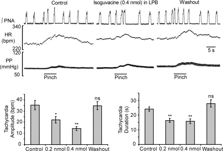
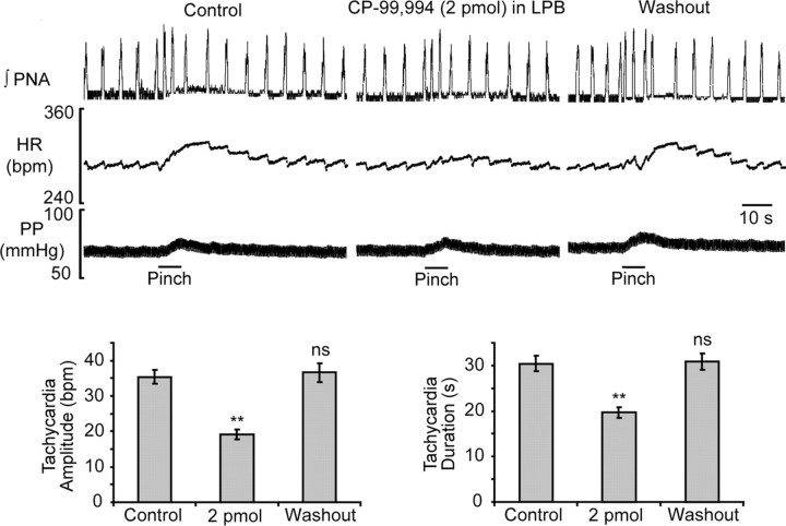
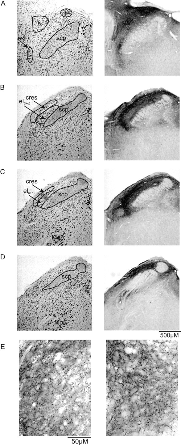
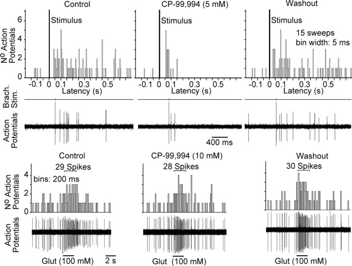
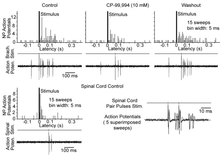
We wanted to ascertain whether the lateral parabrachial nucleus was involved in mediating the heart-rate response evoked during stimulation of somatic nociceptors. Reversible inactivation of the lateral parabrachial nucleus, using a GABA(A) agonist, reduced the reflex tachycardia evoked during noxious (mechanical) stimulation of the forelimb by approximately 50%. The same effect was observed after blockade of neurokinin 1 receptors within the lateral parabrachial nucleus, indicating a possible involvement for substance P as a neurotransmitter. Immunocytochemistry revealed a strong expression of substance P-immunoreactive fibers and boutons in all lateral subnuclei, but they were particularly dense in the lateral crescent subnucleus. Histological verification showed that the most effective injection sites for attenuating the noxious-evoked tachycardia were all placed in or near to the lateral crescent nucleus of the lateral parabrachial complex. Many single units recorded from this region were activated by high-intensity brachial nerve stimulation. The brachial nerve evoked firing responses of some of these neurons was reversibly reduced after local delivery of a neurokinin 1 receptor antagonist. However, only a minority of these neurons followed a paired-pulse stimulation protocol applied to the spinal cord, suggesting a predominance of indirect projections from the spinal cord to the parabrachial nucleus. We conclude that the cardiac component of the response to somatic nociception involves indirect spinal pathways that most likely excite neurons located in the lateral crescent nucleus of the parabrachial complex via activation of neurokinin 1 receptors.


Figures

Figure 1.
Figure 1.
Bilateral microinjection of isoguvacine (a GABAA receptor agonist) into the LPB attenuates the reflex tachycardic response evoked by stimulation of forelimb nociceptors. The attenuation was reversible after a 10 min washout period. In contrast, the reflex changes in respiratory activity and peripheral vascular resistance remained unaffected. *p < 0.05; **p < 0.01; two-way ANOVA and paired two-tailed Student's t test. ns, Not significant.
Figure 3.
Figure 3.
Semi-schematic drawings of coronal sections through the parabrachial complex (A-D, rostral to caudal) illustrating the injection sites for isoguvacine (squares) and for the NK1 receptor antagonist CP-99,994 (circles). To identify the individual pairs of bilateral isoguvacine injection sites, compare the letters A-Q. The pairs of bilateral CP-99,994 injection sites are labeled with numbers 1-7, and extracellular recording sites are indicated by the shaded area. The circles and squares shown in black represent the most effective injection sites in relation to the attenuation of the nociceptive tachycardia (≥50% attenuation). Symbols shown in gray were ineffective or only showed weak attenuation of the cardiac response. Dll, Dorsal nucleus of the lateral lemniscus; me5, mesencephalic tract of the trigeminus; Mo5, motor nucleus of the trigeminus; scp, superior cerebellar peduncle, nuclei of the parabrachial complex; c, central nucleus; cres, lateral crescent nucleus; d, dorsal nucleus; el, external lateral nucleus (eli, inner section; elo, outer section); exl, extreme lateral nucleus; il, internal lateral nucleus; v, ventral nucleus; s, superior nucleus; w, waste nucleus.
Figure 2.
Figure 2.
Bilateral microinjection of CP-99,994 (an NK1 receptor antagonist) into the LPB reversibly attenuates the amplitude and duration of the tachycardia evoked after somatic nociception. Although the tachycardia was attenuated, the changes in respiratory activity and peripheral vascular resistance remained unaffected. **p < 0.01; two-way ANOVA and paired two-tailed Student's t test. ns, Not significant.
Figure 4.
Figure 4.
Series of photomicrographs of coronal sections illustrating the cytoarchitecture of the parabrachial complex with thionin staining (left column) and adjacent sections showing immunoreactivity for substance P (right column). A-D, From rostral to caudal; compare the dense immunoreactivity of the lateral crescent nucleus with the injection sites presented in Figure 3. E is a higher magnification of a substance P-immunoreactive terminal field within the lateral parabrachial nucleus. Note the punctate button-like immunoreactivity surrounding somata (in particular, see top left corner of the right photograph) of the lateral parabrachial complex, suggesting potential SPergeric synaptic contacts in this area. For abbreviations, see Figure 3.
Figure 5.
Figure 5.
Top, Peristimulus time histogram and raw data from a single LPB neuron demonstrating that the NK1 receptor antagonistCP-99,994 attenuated the evoked response during electrical stimulation of the ipsilateral brachial nerve (Brach. Stim.). The effect of CP-99,994 was reversible after a 20 min washout period. Bottom, Glutamate (Glut) applied directly to neurons increased firing, which was unaffected with or without NK1 receptor blockade, indicating integrity of the neurons and an absence of any nonspecific effect of CP-99,994.
Figure 6.
Figure 6.
Peristimulus time histograman draw data from an LPB neuron during electrical stimulation of the ipsilateral brachial nerve (Brach. Stim.) and cervical spinal cord (Spinal Stim.). The synaptic evoked response was sensitive to CP-99,994 in a reversible manner. The spinal input to this neuron was comparable with that evoked from the brachial nerve, albeit at a shorter onset latency. Paired-pulse stimulation with a 10 ms interval of the spinal cord evoked reliable synaptic responses to both stimuli, suggesting a relatively direct synaptic connection from ascending spinal afferents.

Similar articles

Cited by

References

    1. Abram SE, Kostreva DR, Hopp FA, Kampine JP (1983) Cardiovascular responses to noxious radiant heat in anesthetized cats. Am J Physiol 245: R576-R580. - PubMed
    1. al'Absi M, Buchanan TW, Marrero A, Lovallo WR (1999) Sex differences in pain perception and cardiovascular responses in persons with parental history for hypertension. Pain 83: 331-338. - PubMed
    1. Allen GV, Cechetto DF (1992) Functional and anatomical organization of cardiovascular pressor and depressor sites in the lateral hypothalamic area. I. Descending projections. J Comp Neurol 315: 313-332. - PubMed
    1. Bang OU, Lund A (1991) Inhibition of glutamate antagonists, MK-801 and NBQX, of cutaneo-cardiovascular pain reflex in rats. Eur J Pharmacol 203: 133-135. - PubMed
    1. Barr GA (1998) Maturation of the biphasic behavioral and heart rate response in the formalin test. Pharmacol Biochem Behav 60: 329-335. - PubMed

Publication types

MeSH terms

LinkOut - more resources