. 2005 Feb 9;25(6):1481-92.
doi: 10.1523/JNEUROSCI.3523-04.2005.

Kv1 K+ channels control Purkinje cell output to facilitate postsynaptic rebound discharge in deep cerebellar neurons

Affiliations

Kv1 K+ channels control Purkinje cell output to facilitate postsynaptic rebound discharge in deep cerebellar neurons

Bruce E McKay et al. J Neurosci. .

Abstract

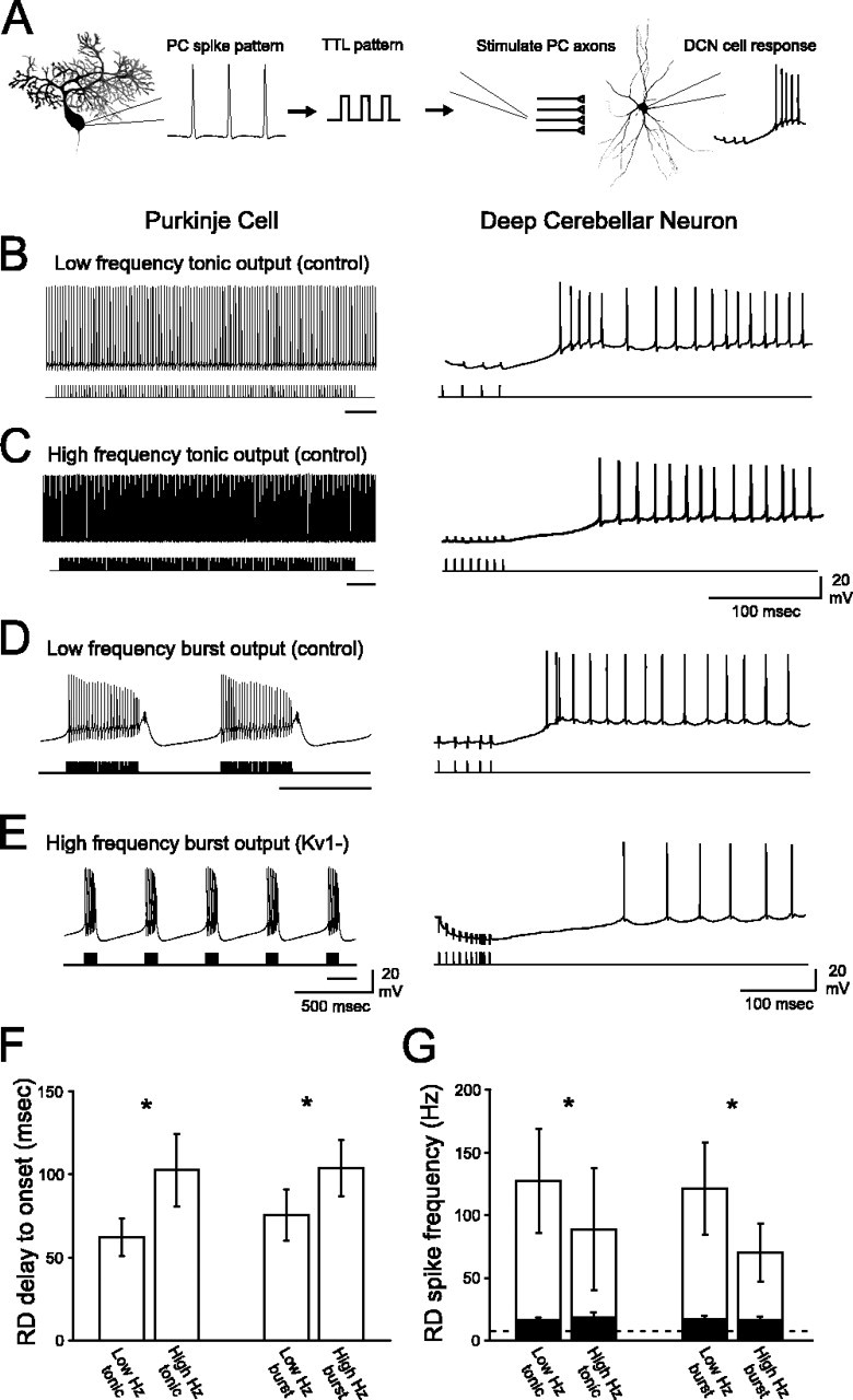
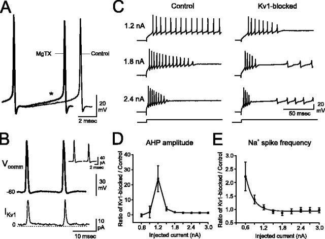
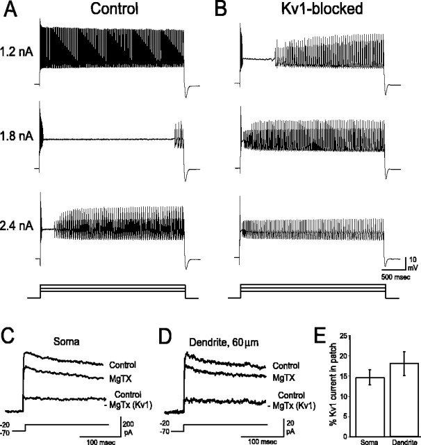
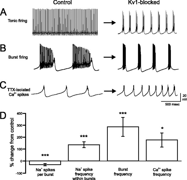
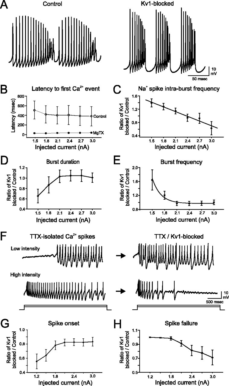
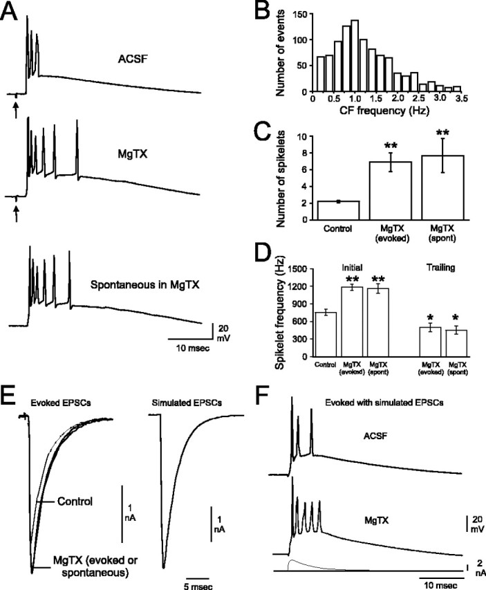
Purkinje cells (PCs) generate the sole output of the cerebellar cortex and govern the timing of action potential discharge from neurons of the deep cerebellar nuclei (DCN). Here, we examine how voltage-gated Kv1 K+ channels shape intrinsically generated and synaptically controlled behaviors of PCs and address how the timing of DCN neuron output is modulated by manipulating PC Kv1 channels. Kv1 channels were studied in cerebellar slices at physiological temperatures with Kv1-specific toxins. Outside-out voltage-clamp recordings indicated that Kv1 channels are present in both somatic and dendritic membranes and are activated by Na+ spike-clamp commands. Whole-cell current-clamp recordings revealed that Kv1 K+ channels maintain low frequencies of Na+ spike and Ca-Na burst output, regulate the duration of plateau potentials, and set the threshold for Ca2+ spike discharge. Kv1 channels shaped the characteristics of climbing fiber (CF) responses evoked by extracellular stimulation or intracellular simulated EPSCs. In the presence of Kv1 toxins, CFs discharged spontaneously at approximately 1 Hz. Finally, "Kv1-intact" and "Kv1-deficient" PC tonic and burst outputs were converted to stimulus protocols and used as patterns to stimulate PC axons and synaptically activate DCN neurons. We found that the Kv1-intact patterns facilitated short-latency and high-frequency DCN neuron rebound discharges, whereas DCN neuron output timing was markedly disrupted by the Kv1-deficient stimulus protocols. Our results suggest that Kv1 K+ channels are critical for regulating the excitability of PCs and CFs and optimize the timing of PC outputs to generate appropriate discharge patterns in postsynaptic DCN neurons.


Figures

Figure 6.
Figure 6.
PC outputs with Kv1 K+ channels intact are optimized to facilitate short latency RDs with high Na+ spike frequency in DCN neurons. A, PC spike outputs were converted to TTL pulse patterns and used as extracellular stimulus patterns to synaptically activate DCN neurons. B-E, Left panels depict spike patterns recorded from PCs, and right panels depict the corresponding DCN neuron responses to the stimulation of inhibitory inputs using each PC discharge as a stimulus template (stimulus artifacts truncated). The black bar under each stimulus protocol corresponds to the expanded region illustrated in the right panels. B, Low-frequency Kv1-intact tonic stimulation of DCN neurons evokes a short-latency RD with high-frequency Na+ spikes. C, Na+ spike frequency decreases, and the delay to RD increases, after high-frequency tonic stimulation (same cell as in B; basal discharge rate, 5.1 Hz). D, The low-frequency Kv1-intact burst stimulation protocol facilitates the generation of a short-latency RD with a high rate of Na+ spike discharge. E, The high-frequency Kv1-deficient burst protocol results in a long latency to elicit RD and low-frequency Na+ spikes during the RD (same cell as in D; basal discharge rate, 6.4 Hz). F, The delay between cessation of inhibitory stimulation and onset of the first Na+ spike in the RD is significantly shorter for both low-frequency stimulus patterns compared with both high-frequency stimulus patterns. G, At the termination of all four stimulus protocols, there was an increase in firing frequency relative to baseline rates of discharge (dashed line; ∼10 Hz) for cells exhibiting high-frequency RDs (unfilled bars) and low-frequency RDs (filled bars). However, pattern-specific differences were noted only for the high-frequency RD group (asterisks). Within the high-frequency RD group, low-frequency tonic and burst stimuli (Kv1-intact) evoked a significantly greater increase in firing rate compared with high-frequency tonic and burst (Kv1-deficient) protocols. Given the large variability in RD spike frequency, the asterisks in G correspond to statistical analyses completed on this data after a log10 transformation.*p < 0.05 (low vs high frequency). Error bars represent SEM.
Figure 3.
Figure 3.
Kv1 K+ channels are activated by Na+ spike-clamp commands and enable sustained low-frequency Na+ spike generation at threshold stimulation intensities. A, Superimposing Na+ spikes from control conditions (black) or after MgTX application (gray) indicates that individual spike properties are not changed when Kv1 K+ channels are blocked (I = 0.9 nA). However, the rate of depolarization during the interspike interval (asterisk) is enhanced in MgTX, leading to higher-frequency spike output. B, Na+ spike-clamp commands (Vcomm) rapidly activate Kv1 currents that deactivate throughout the interspike interval (IKv1). Inset depicts the current activated by a single spike within the spike-clamp command under control (left) and Kv1-blocked (right) conditions. C, In control aCSF and from a holding potential of -70 mV, low-intensity current injection (e.g., 1.2 nA) evokes a train of Na+ action potentials. Higher-intensity stimulation (e.g., 1.8 nA) leads to Na+ spike failure and the emergence of a plateau potential. Additional stimulation (e.g., 2.4 nA) results in rapid Na+ spike failure and the appearance of Ca2+ spikes (C) and Ca-Na bursts (data not shown; see Fig. 1 A). Blockade of Kv1 K+ channels increases the frequency of spike output and results in a failure of spike generation early in the current step. The current injection threshold required to evoke both the plateau potential as well as Ca-Na bursts or Ca2+ spikes is lower. With greater stimulation (e.g., 1.8 or 2.4 nA), Na+ spikes fail rapidly, the duration of the plateau potential is minimal, and Ca-Na bursts or Ca2+ spikes appear very early in the current step. D, The rate at which AHP amplitude decreases throughout a train of Na+ spikes reveals that Kv1K+ channels function to maintain cell output over a narrow window of current injection intensities. The rate of change in AHP amplitude during toxin application was normalized to the rate of change during control conditions. E, Blockade of Kv1 K+ currents results in a marked increase in Na+ spike frequency for threshold levels of current injection. n = 7-13 cells per data point for D and E. Error bars represent SEM.
Figure 1.
Figure 1.
Kv1 K+ channels regulate PC output. A, From a holding potential of -70 mV, a low-intensity current injection (1.2 nA; 3000 ms) evokes a train of high-frequency Na+ action potentials. With progressively greater stimulation (1.8 nA), Na+ spikes are driven to inactivation. A long plateau depolarization is uncovered, terminating with the appearance of Ca2+ spikes and Ca-Na bursts. With intense stimulation (2.4 nA), Na+ spikes inactivate rapidly, the duration of the plateau potential is shortened, and Ca-Na bursts appear early in the record. B, In the presence of 100 nm MgTX, Na+ spikes inactivate rapidly even with low-intensity stimulation (1.2 nA). The current injection threshold to evoke Ca2+ spikes and Ca-Na bursts substantially decreases and is accompanied by a pronounced shortening of the plateau potential before Ca-Na burst onset. C, D, Outside-out patches extracted from somatic (C) and dendritic (D) membranes contain MgTX-sensitive Kv1 K+ currents (steps from -70 to -20 mV). Difference currents (control record - MgTX record) reveal that PC Kv1 K+ currents are noninactivating. E, Kv1 K+ currents account for ∼15% of the total K+ current in outside-out patches sampled from both somatic (n = 9) and dendritic (n = 5) membranes. Error bars represent SEM.
Figure 2.
Figure 2.
Kv1 K+ channels regulate output of spontaneously active PCs. A, B, Under normal conditions, PCs generate either continuous trains of fast Na+ action potentials (A) or fire with the characteristic trimodal pattern that includes a low-frequency Ca-Na burst output (B). Blockade of Kv1 K+ channels converts both tonic Na+ spike and Ca-Na burst output PC types to a high-frequency Ca-Na burst mode (A, B). The high-frequency bursts evoked by toxin application were comparable between tonic (A) and burst-firing (B) cells; the toxin-evoked bursts illustrated in the figure reflect the range of burst frequencies that could result from toxin application. C, TTX application isolated the Ca2+ spike underlying each Ca-Na burst. MgTX increased the frequency of spontaneous Ca2+ spike output dramatically and to a level similar to the frequency of Ca-Na bursts. D, Without Kv1 K+ channels, the number of Na+ spikes within each Ca-Na burst decreases, and the frequency of Na+ spikes within each Ca-Na burst increases. The frequencies of spontaneous Ca-Na bursts and TTX-isolated Ca2+ spike outputs are comparably increased after Kv1 blockade.*p < 0.05; ***p < 0.001. Error bars represent SEM.
Figure 4.
Figure 4.
Kv1 K+ channels control Ca-Na burst output and Ca2+ spike generation in PCs. A, Blockade of Kv1 K+ channels decreases the number and increases the frequency of Na+ spikes fired within individual bursts (I = 1.5 nA). The burst output frequency also increases. B, Plateau potential duration is significantly shortened after Kv1K+ channel blockade. C, The frequency of Na+ spike generation within bursts is accelerated by Kv1 blockade at low stimulus intensities. D, E, Burst duration (D) is substantially shorter at near burst-threshold stimulation intensities, whereas burst frequency (E) markedly increases over this stimulus intensity range. F-H, In the presence of TTX, low-intensity depolarizing current (0.9 nA) evokes a plateau potential followed by Ca2+ spike generation (F). The time of Ca2+ spike onset, normalized to control latencies, is substantially shorter after Kv1 blockade (G). With higher-intensity stimulation (2.4 nA), TTX-isolated Ca2+ spikes are evoked throughout the entire current step with a moderate reduction in frequency toward the end of the step (F). Without Kv1 K+ channels, sustained generation of Ca2+ spikes during high-intensity stimulation is no longer possible (H). n = 11-20 cells per data point for B; n = 9-11 cells per data point for C-E; n = 7-9 cells per data point for G and H. Error bars represent SEM.
Figure 5.
Figure 5.
Kv1 K+ channels modulate PC responses to CF discharge and regulate the presynaptic excitability of CFs. A, Evoked CF responses are markedly changed after Kv1 blockade (arrows denote stimulus artifacts). Although never seen in control solutions, spontaneous CF discharge is elicited by application of Kv1 K+ channel blockers (bottom trace). B, Spontaneous discharges of CFs in MgTX occur over a broad range of frequencies with a peak at ∼1.2 Hz (total, 1005 events plotted). The distribution shown has been truncated at 3.5 Hz, which removed ∼7% of the data that fell outside of a normal distribution. These outlying events were evenly distributed between 3.5 and 13 Hz. C, The number of CF spikelets increases approximately threefold during toxin application. D, The frequency of the initial (2) CF spikelets is increased during Kv1 blockade. Trailing spikelets (an additional 2-7 spikelets per CF event) seen only during toxin application are generated at a significantly lower frequency than the initial spikelets. Note that the characteristics of evoked and spontaneous CF events in MgTX are identical. E, CF EPSCs evoked in control solutions or in the presence of MgTX (superimposed in left panel). The kinetics of evoked EPSCs were used to generate simulated EPSC waveforms (right panel). F, Simulated control CF EPSCs (bottom trace) delivered in current-clamp mode result in CF-like events. Blockade of Kv1 K+ channels results in increased spikelet generation in response to a simulated control EPSC waveform (depicted beneath the spike responses).*p < 0.05 and **p < 0.01 compared with control. Error bars represent SEM.

Similar articles

Cited by

References

    1. Aizenman CD, Linden DJ (1999) Regulation of the rebound depolarization and spontaneous firing patterns of deep nuclear neurons in slices of rat cerebellum. J Neurophysiol 82: 1697-1709. - PubMed
    1. Aizenman CD, Manis PB, Linden DJ (1998) Polarity of long-term synaptic gain change is related to postsynaptic spike firing at a cerebellar inhibitory synapse. Neuron 21: 827-835. - PubMed
    1. Albus JS (1971) A theory of cerebellar function. Math Biosci 10: 25-61.
    1. Bekkers JM, Delaney AJ (2001) Modulation of excitability by α-dendrotoxin-sensitive potassium channels in neocortical pyramidal neurons. J Neurosci 21: 6553-6560. - PMC - PubMed
    1. Brew HM, Forsythe ID (1995) Two voltage-dependent K+ conductances with complementary functions in postsynaptic integration at a central auditory synapse. J Neurosci 15: 8011-8022. - PMC - PubMed

Publication types

MeSH terms

LinkOut - more resources