

Amygdalar inactivation blocks stress-induced impairments in hippocampal long-term potentiation and spatial memory
- PMID: 15703407
- PMCID: PMC6725997
- DOI: 10.1523/JNEUROSCI.4623-04.2005
Amygdalar inactivation blocks stress-induced impairments in hippocampal long-term potentiation and spatial memory
Abstract
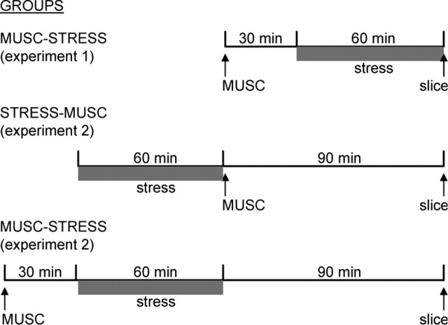
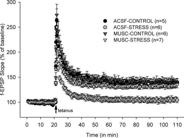
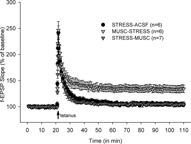
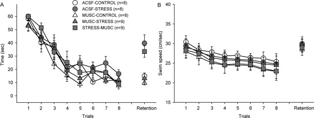
Electrolytic lesions to the amygdala, a limbic structure implicated in stress-related behaviors and memory modulation, have been shown to prevent stress-induced impairments of hippocampal long-term potentiation (LTP) and spatial memory in rats. The present study investigated the role of intrinsic amygdalar neurons in mediating stress effects on the hippocampus by microinfusing the GABA(A) receptor agonist muscimol into the amygdala and examining stress effects on Schaffer collateral/commissural-CA1 LTP and spatial memory. The critical period of the amygdalar contribution to stress effects on hippocampal functions was determined by applying muscimol either before stress or immediately after stress. Our results indicate that intra-amygdalar muscimol infusions before uncontrollable restraint-tailshock stress effectively blocked stress-induced physiological and behavioral effects. Specifically, hippocampal slices prepared from vehicle-infused stressed animals exhibited markedly impaired LTP, whereas slices obtained from muscimol-infused stressed animals demonstrated robust LTP comparable with that of unstressed animals. Correspondingly, vehicle-infused stressed animals displayed impaired spatial memory (on a hidden platform version of the Morris water maze task), whereas muscimol-infused stressed animals revealed unimpaired spatial memory. In contrast to prestress muscimol effects, however, immediate poststress infusions of muscimol into the amygdala failed to interfere with stress impairments of LTP and spatial memory. Together, these results suggest that the amygdalar neuronal activity during stress, but not shortly after stress, is essential for the emergence of stress-induced alterations in hippocampal LTP and memory.
Figures
References
-
- Abe K (2001) Modulation of hippocampal long-term potentiation by the amygdala: a synaptic mechanism linking emotion and memory. Jpn J Pharmacol 86: 18-22. - PubMed
-
- Adamec RE, Burton P, Shallow T, Budgell J (1999) Unilateral block of NMDA receptors in the amygdala prevents predator stress-induced lasting increases in anxiety-like behavior and unconditioned startle—effective hemisphere depends on the behavior. Physiol Behav 65: 739-751. - PubMed
-
- Aggleton JP (1986) A description of the amygdalo-hippocampal interconnections in the macaque monkey. Exp Brain Res 64: 515-526. - PubMed
-
- Anderson JW (1954) The production of ultrasonic sounds by laboratory rats and other mammals. Science 119: 808-809. - PubMed
Publication types
MeSH terms
Substances
Grants and funding
LinkOut - more resources
Full Text Sources
Medical
Miscellaneous