. 2005 Apr;11(4):470-86.
doi: 10.1261/rna.7215305. Epub 2005 Feb 9.

Binding of eukaryotic initiation factor 3 to ribosomal 40S subunits and its role in ribosomal dissociation and anti-association

Affiliations

Binding of eukaryotic initiation factor 3 to ribosomal 40S subunits and its role in ribosomal dissociation and anti-association

Victoria G Kolupaeva et al. RNA. 2005 Apr.

Abstract

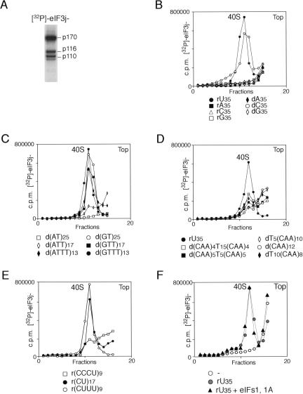
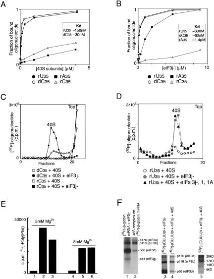
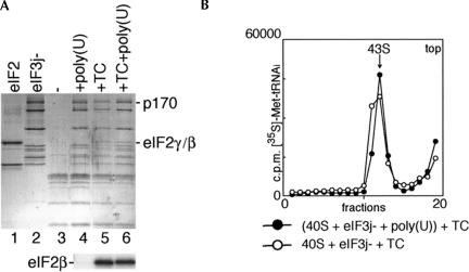
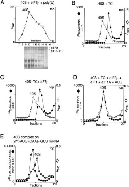
The multisubunit eukaryotic initiation factor (eIF) 3 plays various roles in translation initiation that all involve interaction with 40S ribosomal subunits. eIF3 can be purified in two forms: with or without the loosely associated eIF3j subunit (eIF3j+ and eIF3j-, respectively). Although unlike eIF3j+, eIF3j- does not bind 40S subunits stably enough to withstand sucrose density gradient centrifugation, we found that in addition to the known stabilization of the eIF3/40S subunit interaction by the eIF2*GTP*Met-tRNA(i)Met ternary complex, eIF3j-/40S subunit complexes were also stabilized by single-stranded RNA or DNA cofactors that were at least 25 nt long and could be flanked by stable hairpins. Of all homopolymers, oligo(rU), oligo(dT), and oligo(dC) stimulated the eIF3/40S subunit interaction, whereas oligo(rA), oligo(rG), oligo(rC), oligo(dA), and oligo(dG) did not. Oligo(U) or oligo(dT) sequences interspersed by other bases also promoted this interaction. The ability of oligonucleotides to stimulate eIF3/40S subunit association correlated with their ability to bind to the 40S subunit, most likely to its mRNA-binding cleft. Although eIF3j+ could bind directly to 40S subunits, neither eIF3j- nor eIF3j+ alone was able to dissociate 80S ribosomes or protect 40S and 60S subunits from reassociation. Significantly, the dissociation/anti-association activities of both forms of eIF3 became apparent in the presence of either eIF2-ternary complexes or any oligonucleotide cofactor that promoted eIF3/40S subunit interaction. Ribosomal dissociation and anti-association activities of eIF3 were strongly enhanced by eIF1. The potential biological role of stimulation of eIF3/40S subunit interaction by an RNA cofactor in the absence of eIF2-ternary complex is discussed.


Figures

FIGURE 1.
FIGURE 1.
Subunit composition of HeLa eIF3j+ and eIF3j− analyzed by electrophoresis on 4%–12% Bis-Tris NuPAGE gel using MOPS buffer system (Invitrogen), followed by Coomassie staining (A). Presence of eIF3 in the peak fraction corresponding to 40S ribosomal subunits after sucrose density gradient centrifugation, analyzed by electrophoresis on (B,D,F) 4%–12% Bis-Tris NuPAGE gel using MES buffer system (Invitrogen) or (C) SDS-11% polyacrylamide, followed by Coomassie staining. (B,F) 40S subunits (lane 7) and eIF3j− (lane 8) and (D) 40S subunits (lane 6) and eIF3j− (lane 7) were incubated in the absence (lane 1) or in the presence of poly- or oligonucleotides, as indicated, and separated by centrifugation in 10%–30% linear sucrose density gradients. (C) 40S subunits were incubated with eIF3j− alone (lane 1), eIF3j−and poly(U) (lane 2), eIF3j− and globin mRNA (lane 3) or eIF3j−, globin mRNA, eIFs 4A, 4B, and 4F (lane 4) and separated by sucrose density gradient centrifugation. eIF3 subunits are labeled to the right of panels A–D and F. (E) Sequences and structures of 5′ stem-U31 and 5′stem-dT40 oligonucleotides.
FIGURE 2.
FIGURE 2.
Activity of RNA and DNA oligonucleotides in promoting association of [32P]-labeled eIF3j− and 40S subunits. (A) [32P]-phosphorylation of eIF3a (p170), eIF3b (p116), and eIF3c (p110) subunits of eIF3j− by the catalytic subunit of cAMP-dependent protein kinase. [32P]-phosphorylated eIF3j− was resolved by gel electrophoresis on 4%–12% Bis-Tris NuPAGE gel using MES buffer system and visualized by autoradiography. (B–F) Binding of [32P]-labeled eIF3j− to 40S subunits in the presence of oligonucleotides and eIF1 and eIF1A, as indicated. Ribosomal complexes were separated by centrifugation in 10%–30% linear sucrose gradients, and aliquots of gradient fractions were analyzed by scintillation counting. The position of 40S subunits determined by optical density is indicated. Sedimentation was from right to left. Upper fractions from the gradient have been omitted for clarity.
FIGURE 3.
FIGURE 3.
Binding of oligonucleotides to eIF3j− and to 40S subunits. (A,B) Affinity measurements made in filter-binding experiments using (A) 40S subunits and (B) eIF3j− with [32P]-labeled oligonucleotides as indicated. The fraction of bound oligonucleotide is the ratio of [32P]-labeled oligonucleotide retained on the filter to the input. Dissociation constants are shown in the inset boxes. (C) Binding of [32P]-labeled oligonucleotide (dC35 or rC35) to 40S subunits in the presence and absence of eIF3j−. (D) Influence of eIF1 and eIF1A on binding of [32P]-labeled rU35 to the 40S subunit in the presence of eIF3j−. Ribosomal complexes were separated by centrifugation in 10%–30% linear sucrose gradients. Sedimentation was from right to left. The position of 40S subunits determined by optical density is indicated. Upper fractions from the gradient have been omitted for clarity. (E) Template-dependent [3H]-polyphenylalanine synthesis by ribosomes assembled by incubation at 5 mM Mg2+ (lanes 1–3) or 8 mM Mg2+ (lanes 4–6) of 40S and 60S subunits without poly(U) (lanes 1,4), 60S subunits with preassembled 40S subunit/poly(U) complexes (lanes 2,5) or poly(U) with preassembled 80S ribosomes (lanes 3,6). All reaction mixtures contained [3H]-Phe-tRNAPhe, EF1A, and EF2. [3H]-polyphenylalanine was counted after TCA precipitation of reaction mixtures on nitro-cellulose filters. (F) UV-crosslinking of [32P]-β-globin mRNA (lanes 1,2) and [32P]-(CUUU)9 (lanes 3–5) to eIF3j− (lanes 1–4) and 40S subunits (lane 5) in binary eIF3j−/RNA complexes (lanes 1,3), 48S complexes (lane 2), and eIF3j−/(CUUU)9/40S subunit complexes (lanes 4,5). Polypeptides resolved by gel electrophoresis were visualized by autoradiography. eIF3a (p170), eIF3b (p116), eIF3d (p66), and eIF3g (p44) are indicated to the right of lanes 2,4. The positions of molecular weight markers are shown to the right of the radiolabeled ribosomal proteins in lane 5.
FIGURE 4.
FIGURE 4.
Binding of eIF2-ternary complex (TC) and eIF3j− to 40S subunits in the presence and absence of poly(U) RNA. (A) Detection of eIF2 and eIF3j− in ribosomal complexes isolated from sucrose density gradients. eIF2 (lane 1), eIF3j− (lane 2), 40S subunits (lane 3), 40S/eIF3j− binary complexes formed in the presence of poly(U) (lane 4), 43S ribosomal complexes formed from purified TC and preassembled eIF3j−/poly(U)/40S subunit complexes (lane 6), and 43S complexes assembled using TC, eIF3j− and 40S subunits (lane 5) were analyzed by gel electrophoresis followed by Coomassie staining (lanes 1–6 of the upper panel) or Western blotting with eIF2βantibodies (lanes 4–6 of the lower panel). eIF3a (p170 subunit) and eIF2β,γ are labeled to the right of the upper panel; eIF2β is labeled to the left of the lower panel. (B) Incorporation of aminoacylated [35S]Met-tRNAiMet into 43S complexes, assembled either from TC and preassembled eIF3j−/poly(U)/40S subunit complexes or from 40S subunits, eIF3j−, and TC. Ribosomal complexes were separated by centrifugation in 10%–30% linear sucrose gradients. Sedimentation was from right to left. The position of 43S complexes is indicated. Upper fractions from the gradient have been omitted for clarity.
FIGURE 5.
FIGURE 5.
Ribosomal dissociation activity of eIF3. Dissociation activity of eIF3j− in the presence of (A) poly(U), (C) eIF2-ternary complex (TC) and (D) poly(U), eIF1, and eIF1A. (B) Dissociation activity of eIF3j+ in the presence of poly(U), eIF1, and eIF1A. (E,F) Resistance of 80S ribosomes to dissociation by (E) eIF1 or eIF1A alone and (F) eIF3c (p110 subunit) alone, with eIF1 or with eIF1 and poly(U). The optical density of ribosomal profiles was measured after centrifugation through 10%–30% linear sucrose gradients. Sedimentation was from right to left. Upper fractions from gradients have been omitted for clarity. (G) Dissociation by eIF3j− (in the presence and absence of poly(U)) of 80S complexes assembled on [32P]-CAAn-GUS mRNA and purified by sucrose density gradient centrifugation. After incubation with eIF3j− and poly(U) as indicated, ribosomal complexes were again centrifuged through 10%–30% linear sucrose gradients. Sedimentation was from right to left. Upper fractions from the gradient have been omitted for clarity.
FIGURE 6.
FIGURE 6.
Mobility of ribosomal complexes during centrifugation through 10%–30% linear sucrose density gradients. (A) Fractionation by sucrose density gradient centrifugation of eIF3j−/poly(U)/40S subunit complexes formed with excess 40S subunits. The positions of 40S subunits and their complexes with eIF3j− were determined by optical density. Polypeptides in different fractions were resolved by electrophoresis followed by Coomassie staining. eIF3a (p170) and eIF3b/3c (p116/p110) are labeled to the right of the lower panel. (B,C) Mobility of preinitiation 43S complexes (consisting of 40S subunits and eIF2-ternary complex [TC]) with (C) or without (B) eIF3, compared to free 40S subunits. (D) Mobility of 43S preinitiation complexes (consisting of 40S subunits, eIF1, eIF1A, TC, eIF3, and AUG triplets) compared to free 40S subunits. The positions of 40S subunits and their complexes with TC, eIF3, eIF1, and eIF1A were determined by optical density and by scintillation counting of [35S]Met-tRNAiMet (B,C,D). (E) Mobility of 48S initiation complexes assembled on [32P]-2-nt-AUG-(CAA)n-GUS mRNA compared to free 40S subunits. The positions of 40S subunits and 48S complexes were determined by optical density and by Cherenkov counting of [32P]-2-nt-AUG-(CAA)n-GUS mRNA. Sedimentation was from right to left. Upper fractions from the gradient have been omitted for clarity.
FIGURE 7.
FIGURE 7.
(A–G) Stimulation of the binding of the eIF2•GTP•Met-tRNAiMet ternary complex (TC) to 40S subunits by other initiation factors and AUG triplets, as indicated. Ribosomal complexes were fractionated by centrifugation in 10%–30% linear sucrose gradients and analyzed by scintillation counting of [35S]Met-tRNAiMet. Sedimentation was from right to left. Upper fractions from each gradient have been omitted for clarity.

Similar articles

Cited by

References

    1. Adler, A., Grossmann, L., and Fasman, G.D. 1967. Single-stranded oligomers and polymers of cytidylic and 2′-deoxycytidylic acids: Comparative optical rotatory studies. Proc. Natl. Acad. Sci. 57: 423–430. - PMC - PubMed
    1. Arnott, S., Chandrasekaran, R., and Leslie, A.G. 1976. Structure of the single-stranded polyribonucleotide polycytidylic acid. J. Mol. Biol. 106: 735–748. - PubMed
    1. Asano, K., Kinzy, T.G., Merrick, W.C., and Hershey, J.W.B. 1997. Conservation and diversity of eukaryotic translation initiation factor eIF3. J. Biol. Chem. 272: 1101–1109. - PubMed
    1. Asano, K., Clayton, J., Shalev, A., and Hinnebusch, A.G. 2000. A multifactor complex of eukaryotic initiation factors, eIF1, eIF2, eIF3, eIF5 and initiator tRNA(Met) is an important translation initiation intermediate in vivo. Genes & Dev. 14: 2534–2546. - PMC - PubMed
    1. Bandyopadhyay, A. and Maitra, U. 1999. Cloning and characterization of the p42 subunit of mammalian translation initiation factor 3 (eIF3): Demonstration that eIF3 interacts with eIF5 in mammalian cells. Nucleic Acids Res. 27: 1331–1337. - PMC - PubMed

Publication types

MeSH terms