. 2005 Apr;11(4):371-82.
doi: 10.1261/rna.7258505. Epub 2005 Feb 9.

Processing bodies require RNA for assembly and contain nontranslating mRNAs

Affiliations

Processing bodies require RNA for assembly and contain nontranslating mRNAs

Daniela Teixeira et al. RNA. 2005 Apr.

Abstract

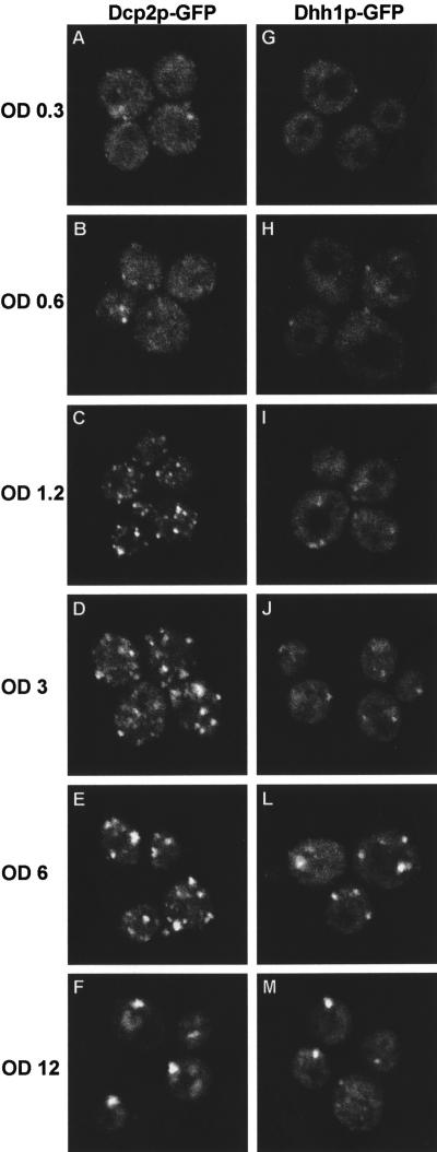
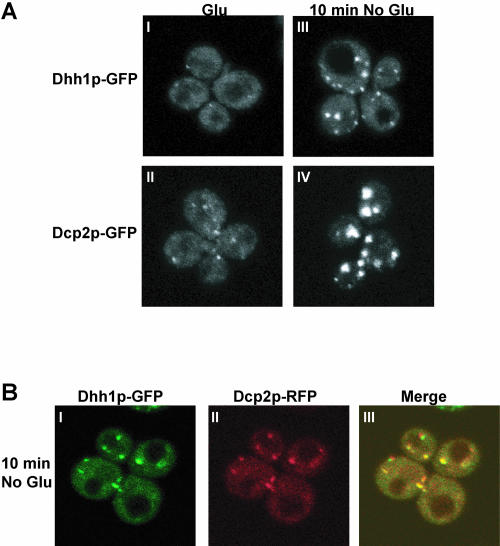
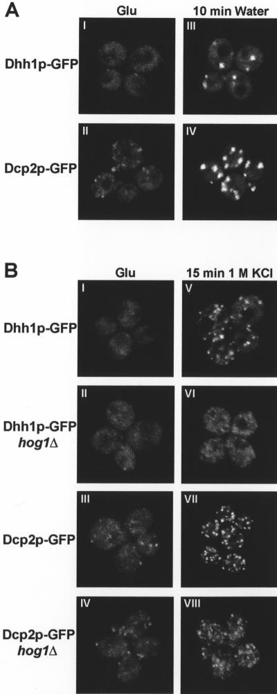
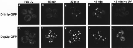
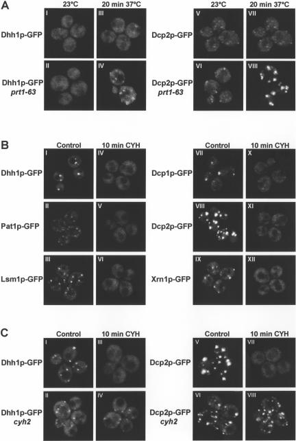
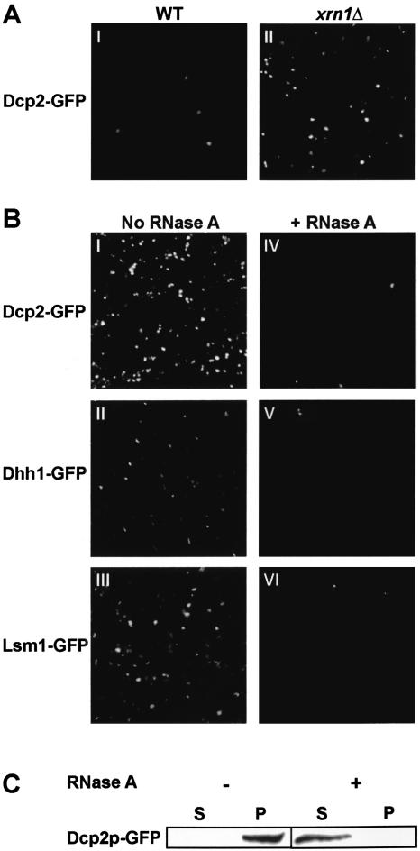
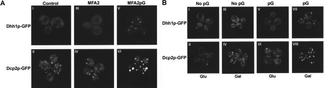
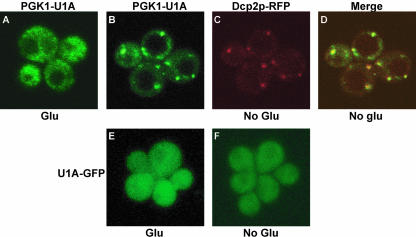
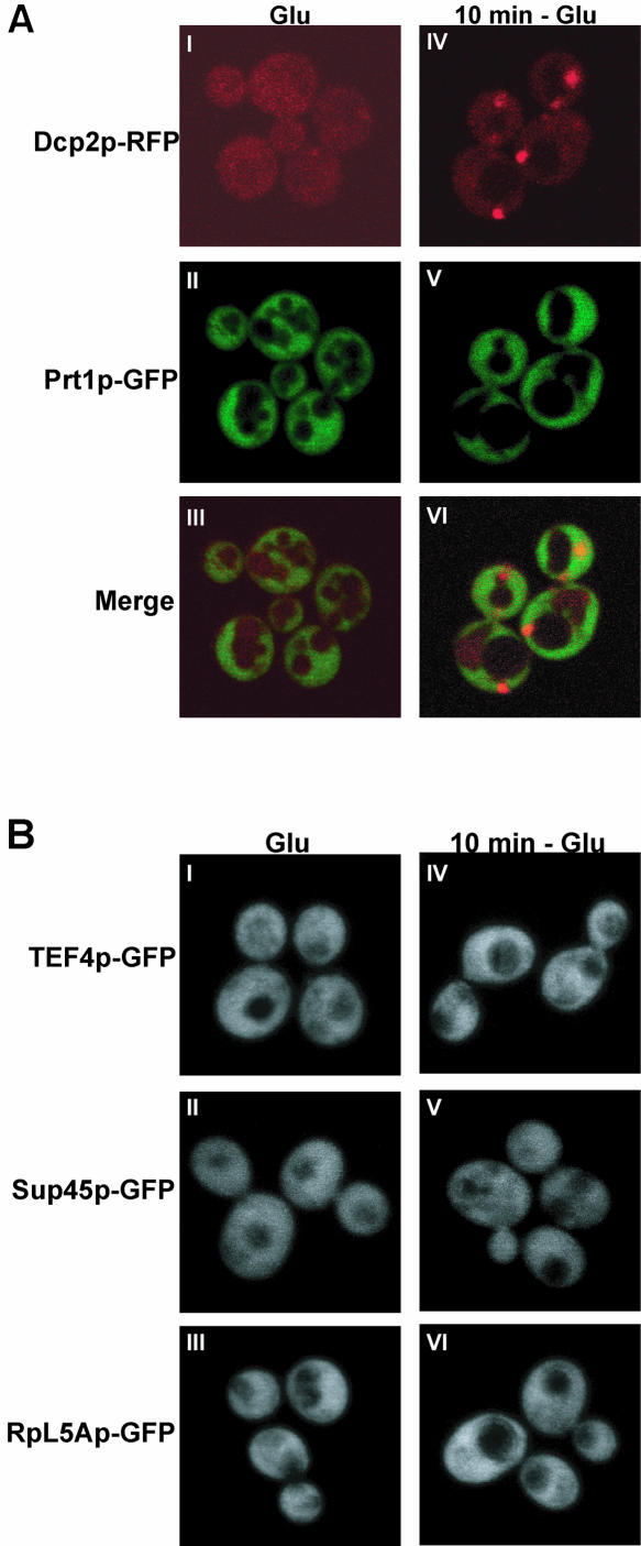
Recent experiments have defined cytoplasmic foci, referred to as processing bodies (P-bodies), wherein mRNA decay factors are concentrated and where mRNA decay can occur. However, the physical nature of P-bodies, their relationship to translation, and possible roles of P-bodies in cellular responses remain unclear. We describe four properties of yeast P-bodies that indicate that P-bodies are dynamic structures that contain nontranslating mRNAs and function during cellular responses to stress. First, in vivo and in vitro analysis indicates that P-bodies are dependent on RNA for their formation. Second, the number and size of P-bodies vary in response to glucose deprivation, osmotic stress, exposure to ultraviolet light, and the stage of cell growth. Third, P-bodies vary with the status of the cellular translation machinery. Inhibition of translation initiation by mutations, or cellular stress, results in increased P-bodies. In contrast, inhibition of translation elongation, thereby trapping the mRNA in polysomes, leads to dissociation of P-bodies. Fourth, multiple translation factors and ribosomal proteins are lacking from P-bodies. These results suggest additional biological roles of P-bodies in addition to being sites of mRNA degradation.


Figures

FIGURE 1.
FIGURE 1.
P-bodies increase with the stage of cell growth. Cells were grown in YPGlu and observed at different stages of cellular growth, as described. The different OD600 units (OD) are indicated at the left side of the figure. Cells expressing a GFP-tagged version of (left panel) Dcp2p (yRP1727) and (right panel) Dhh1p (yRP1724) are shown.
FIGURE 2.
FIGURE 2.
P-bodies size increases under glucose deprivation. (A) Cells were grown in YPGlu, washed in YP, and resuspended in YP for 10 min before being collected. For observation, cells were washed with SC with (I, II) or without (III, IV) Glu, resuspended in the same SC, respectively, and observed. Cells expressing a GFP-tagged version of (top panel) Dhh1p (yRP1724) and (bottom panel) Dcp2p (yRP1727) are shown. (B) Cells were grown in SC plus Glu, washed, and resuspended in SC for 10 min before being collected. For observation, cells were washed and resuspended in SC without Glu. Cells coexpressing (I) a GFP-tagged version of Dhh1p (yRP1724) and (II) a RFP-tagged version of Dcp2p (pRP1186) are shown. (III) A merged view is shown.
FIGURE 3.
FIGURE 3.
P-bodies are increased by osmotic stress. (A) The cells were grown in YPGlu, washed, and resuspended in water for 10 min before collection. For observation, cells were washed and resuspended in water (III, IV). Cells expressing a GFP-tagged version of (top panel) Dhh1p (yRP1724) and (bottom panel) Dcp2p (yRP1727) are shown. (B) Cells expressing a GFP-tagged version of Dhh1p in a wild-type (yRP1724) or hog1Δ (yRP1912) strain (left panel), and Dcp2p in a wild-type (yRP1727) or hog1Δ (yRP1913) strain (right panel) after exposure to 1 M KCl for 15 min (VVIII) are shown. Cells were washed and resuspended in SC plus Glu supplemented or not with the same concentration of KCl, and observed.
FIGURE 4.
FIGURE 4.
P-bodies are increased by exposure to UV light. Localization of GFP-tagged (top panel) Dhh1p (yRP1724) and (bottom panel) Dcp2p (yRP1727). Cells (A,B) before UV treatment and (C,D) 15 min, (E,F) 30 min, and (G,H) 45 min after UV treatment. (I,J) Localization of unexposed cells treated the same way as UV-treated cells.
FIGURE 5.
FIGURE 5.
(A) P-bodies are affected by translation initiation rates. Cells expressing a GFP-tagged version of Dhh1p carrying the conditional ts allele prt1-63 (yRP1826) or a wild-type allele (yRP1724) and cells expressing a GFP-tagged version of Dcp2p carrying the conditional ts allele prt1-63 (yRP1827) or a wild-type allele (yRP1727) were grown in YPGlu at 23°C (I,II,V,VI) and then shifted to 37°C for 20 min (III,IV,VII,VIII). Cells were collected, washed, and resuspended in SC with Glu; cells were kept at 37°C for observation. (B) Inhibition of translation elongation disrupts P-bodies. Cells were grown in YPGlu. The culture was divided into two samples; cycloheximide (CYH) at a concentration of 100 μg/mL was added to one sample. The cells were grown for another 10 min before they were collected. The water used for washing and resuspension of the cycloheximide-treated cells contained the same concentration of the drug (IV–VI,XXII). The other sample was treated similarly but without adding the drug (I–III,VII–IX). Dcp1p (yRP1726), Dcp2p (yRP1727), Dhh1p (yRP1724), Lsm1p (yRP1729), Pat1p (yRP1728), and Xrn1p (yRP1730) GFP-tagged version proteins are shown. (C) Inhibition of translation elongation in a strain altered in ribosomal protein L28 does not affect P-bodies. Cells were grown in YPGlu, the culture was divided into two samples; cycloheximide at a concentration of 2 μg/mL for 10 min was added to one sample. The cells were washed in water with (III,IV,VII,VIII) or without (I,II,V,VI) cycloheximide before they were collected and observed.
FIGURE 6.
FIGURE 6.
P-bodies are sensitive to RNase A digestion. (A) Microscopic analysis of P-body concentrated pellet from a (I) wild-type (yRP1727) and an (II) xrn1Δ (yRP1923) strain expressing a GFP-tagged version of Dcp2p. (B) Fluorescence microscopy of the P-body concentrated fractions in the presence (IVVI) and absence (IIII) of RNase A. Dcp2p (yRP1923), Dhh1p (yRP1738), Lsm1p (yRP1924) GFP-tagged version proteins in xrn1Δ strain. (C) Western blot analysis of the supernatant (S) and pellet (P) fractions from a Dcp2p-GFP xrn1Δ (yRP1923) strain in the presence (+) or absence (−) of RNase A.
FIGURE 7.
FIGURE 7.
P-body integrity is dependent on mRNA. (A) Dhh1p GFP-tagged strain (yRP1724) and Dcp2p GFP-tagged strain (yRP1727) cells transformed with (III,IV) pRP1083 [expressing MFA2 mRNA with no functional poly(G) site] or (V,VI) pRP1081 [expressing the MFA2 mRNA with poly(G)]; (I,II) control cells. Cells expressing a GFP-tagged version of (top panel) Dhh1p (yRP1824) and (bottom panel) Dcp2p (yRP1825). (B) Cells lacking the GAL–PGK1-pG/MFA2-pG constructs (I–IV) and expressing a GFP-tagged version of (top panel) Dhh1p (yRP1824) and (bottom panel) Dcp2p (yRP1825) grown in the presence of Glu (I,II) or Gal (III,IV). Cells expressing a GFP-tagged version of (top panel) Dhh1p (yRP1724) and (bottom panel) Dcp2p (yRP1727), containing the PGK1-pG/MFA2-pG constructs (VVIII) grown in the presence of Glu (V,VI) or Gal (VII,VIII).
FIGURE 8.
FIGURE 8.
mRNA localizes to P-bodies during translation repression. Yeast strain yRP684 coexpressing (AC) U1A-GFP (pRP1187), PGK1-U1A (pPS2037), and Dcp2p-RFP (pRP1155), or (E,F) control U1A-GFP (pRP1187), was grown in SC plus Glu. For glucose depletion, cells were washed and resuspended in SC for 10 min before collection. For observation, cells were washed and resuspended in SC with (A,E) or without (B,C,F) Glu. (D) Merged view.
FIGURE 9.
FIGURE 9.
Translation factors are not associated with P-bodies. (A) Cells were grown in SC plus Glu. For glucose depletion, cells were washed in SC for 10 min before collection. For observation, cells were washed and resuspended in SC with (I,II) or without (IVV) Glu. Cells coexpressing (I,II,IV,V) a GFP-tagged version of Prt1p (yRP1828) and an RFP-tagged version of Dcp2p (pRP1186). (III,VI) Merged views. (B) GFP-tagged versions of TEF4 (yRP1829), Sup45p (yRP1830), and RpL5p (yRP1911) were grown in YPGlu and observed as described in Figure 2A. Cells observed in the (IIII) presence or (IVVI) absence of Glu are shown.

Similar articles

Cited by

References

    1. Anderson, P. and Kedersha, N. 2002. Stressful initiations. J. Cell Sci. 115: 3227–3234. - PubMed
    1. Ashe, M.P., De Long, S.K., and Sachs, A.B. 2000. Glucose depletion rapidly inhibits translation initiation in yeast. Mol. Biol. Cell 3: 833–848. - PMC - PubMed
    1. Bashkirov, V.I., Scherthan, H., Solinger, J.A., Buerstedde, J.M., and Heyer, W.D. 1997. A mouse cytoplasmic exoribonuclease (mXRN1p) with preference for G4 tetraplex substrates. J. Cell Biol. 136: 761–773. - PMC - PubMed
    1. Beelman, C.A. and Parker, R. 1994. Differential effects of translational inhibition in cis and in trans on the decay of the unstable yeast MFA2 mRNA. J. Biol. Chem. 13: 9687–9692. - PubMed
    1. Birrell, G.W., Giaever, G., Chu, A.M., Davis, R.W., and Brown, J.M. 2001. A genome-wide screen in Saccharomyces cerevisiae for genes affecting UV radiation sensitivity. Proc. Natl. Acad. Sci. 98: 12608–12613. - PMC - PubMed

MeSH terms