

Glucocorticoids induce G1 cell cycle arrest in human neoplastic thymic epithelial cells
- PMID: 15703942
- PMCID: PMC12161239
- DOI: 10.1007/s00432-004-0646-8
Glucocorticoids induce G1 cell cycle arrest in human neoplastic thymic epithelial cells
Abstract
Purpose: Glucocorticoids exert anti-proliferative effects in various cell types and have long been known to induce apoptosis in thymocytes. Although a few reports have described the regression of human thymoma with glucocorticoid therapy, its effects on neoplastic thymic epithelial cells (TECs) have not been reported. In the present study, we investigated glucocorticoid receptor (GR) expression on neoplastic TECs and the effects of glucocorticoids in vitro on the cell cycle progression of tumor cells.
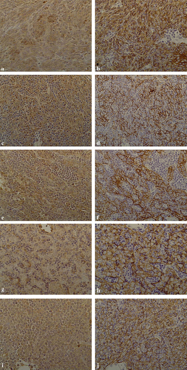
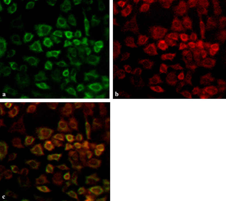
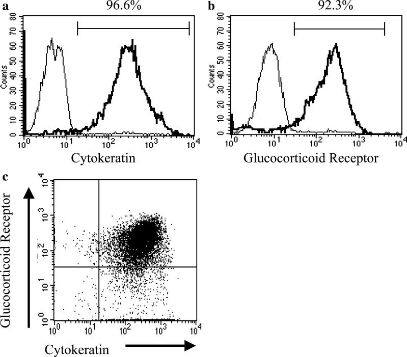
Patients and methods: Thymoma specimens were obtained during surgery from 21 patients. Three of the specimens with glucocorticoid therapy were examined using the TdT-mediated dUTP-biotin nick-end labeling method. Primary tumor specimens from ten untreated thymomas were examined for GR expression by immunohistochemistry. Isolated neoplastic TECs from the remaining eight untreated thymomas were examined using immunohistochemistry, flow cytometric and cell cycle analysis.
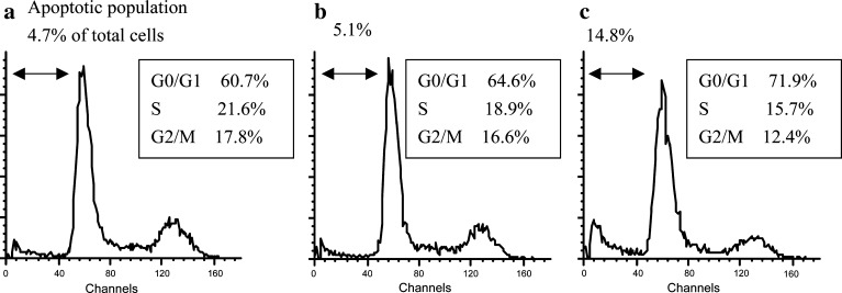
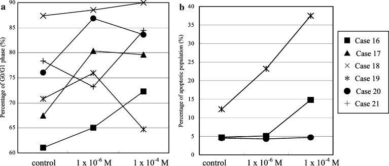
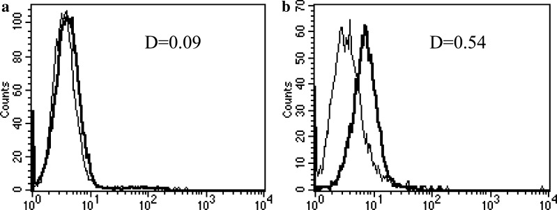
Results: GR are expressed on neoplastic TECs as well as on non-neoplastic thymocytes in thymomas, regardless of WHO histological classification. Glucocorticoids caused an accumulation of TEC in G0/G1 phase in all cases examined (n = 6), and also induced apoptosis in the three with the lowest levels of Bcl-2 expression.
Conclusions: Our results indicate that neoplastic TECs express GR and that glucocorticoids directly suppress their in vitro proliferation.
Figures
Similar articles
-
Characteristics of Good's Syndrome in China: A Systematic Review.Chin Med J (Engl). 2017 Jul 5;130(13):1604-1609. doi: 10.4103/0366-6999.208234. Chin Med J (Engl). 2017. PMID: 28639577 Free PMC article.
-
Thymoma: current diagnosis and treatment.Chin Med J (Engl). 2013;126(11):2186-91. Chin Med J (Engl). 2013. PMID: 23769581
-
Differential Expression Patterns of Bcl-2, D2-40, β-Catenin and E-Cadherin in Thymomas: Correlation with Clinical Stages and Subtypes.Appl Immunohistochem Mol Morphol. 2025 Jul 1;33(4):250-256. doi: 10.1097/PAI.0000000000001264. Epub 2025 May 13. Appl Immunohistochem Mol Morphol. 2025. PMID: 40356427
-
Type B thymomas in patients with myasthenia gravis display a distinctive pattern of αβ TCR and IL-7 receptor α expression on CD4+CD8+ thymocytes.Autoimmunity. 2024 Apr 27;57(1):2347379. doi: 10.1080/08916934.2024.2347379. Epub 2024 May 9. Autoimmunity. 2024. PMID: 38723105
-
Systemic pharmacological treatments for chronic plaque psoriasis: a network meta-analysis.Cochrane Database Syst Rev. 2021 Apr 19;4(4):CD011535. doi: 10.1002/14651858.CD011535.pub4. Cochrane Database Syst Rev. 2021. Update in: Cochrane Database Syst Rev. 2022 May 23;5:CD011535. doi: 10.1002/14651858.CD011535.pub5. PMID: 33871055 Free PMC article. Updated.
Cited by
-
Rapid regression of stage IVb invasive thymoma under palliative corticosteroid administration.Gen Thorac Cardiovasc Surg. 2007 Apr;55(4):180-3. doi: 10.1007/s11748-006-0099-x. Gen Thorac Cardiovasc Surg. 2007. PMID: 17491357
-
Treatment of thymoma with low-dose glucocorticoids before surgery for significant tumor shrinkage: A case report.World J Clin Cases. 2025 Apr 16;13(11):98979. doi: 10.12998/wjcc.v13.i11.98979. World J Clin Cases. 2025. PMID: 40242232 Free PMC article.
-
The Never-Ending History of Octreotide in Thymic Tumors: A Vintage or A Contemporary Drug?Cancers (Basel). 2022 Feb 2;14(3):774. doi: 10.3390/cancers14030774. Cancers (Basel). 2022. PMID: 35159040 Free PMC article. Review.
-
Metastatic Thymoma-Associated Myasthenia Gravis: Favorable Response to Steroid Pulse Therapy Plus Immunosuppressive Agent.Med Sci Monit. 2017 Mar 9;23:1217-1223. doi: 10.12659/msm.902442. Med Sci Monit. 2017. PMID: 28278141 Free PMC article.
-
Single-center evaluation of prognostic factors for thymoma treated by surgery: a retrospective study.J Cardiothorac Surg. 2021 Jan 7;16(1):8. doi: 10.1186/s13019-020-01386-7. J Cardiothorac Surg. 2021. PMID: 33413522 Free PMC article.
References
-
- Auphan N, DiDonato JA, Rosette C, Helmberg A, Karin M (1995) Immunosupression by glucocorticoids: inhibition of NF-κB activity through induction of IκB synthesis. Science 270:286–290 - PubMed
-
- Beato M, Herrlich P, Schutz G (1995) Steroid hormone receptors: many actors in search of a plot. Cell 83:851–857 - PubMed
-
- Berki T, Kumanovics G, Kumanovics A, Falus A, Ujhelyi E, Nemeth P (1998) Production and flow cytometric application of a monoclonal anti-glucocorticoid receptor antibody. J Immunol Methods 214:19–27 - PubMed
-
- Berki T, Palinkas L, Boldizsar F, Nemeth P (2002) Glucocorticoid (GC) sensitivity and GC receptor expression differ in thymocyte subpopulations. Int Immunol 14:463–469 - PubMed
-
- Cohen JJ, Duke RC (1984) Glucocorticoid activation of a calcium-dependent endonuclease in thymocyte nuclei leads to cell death. J Immunol 132:38–42 - PubMed
MeSH terms
Substances
LinkOut - more resources
Full Text Sources
Medical
