

Backbone solution structures of proteins using residual dipolar couplings: application to a novel structural genomics target
- PMID: 15704012
- PMCID: PMC1815388
- DOI: 10.1007/s10969-005-4899-5
Backbone solution structures of proteins using residual dipolar couplings: application to a novel structural genomics target
Abstract
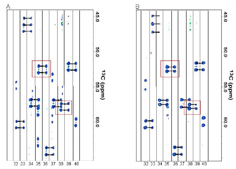
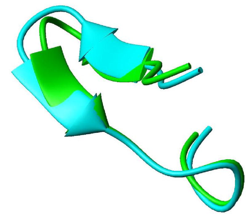
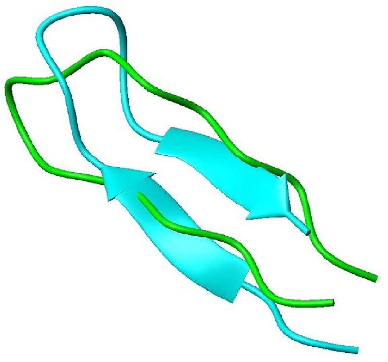
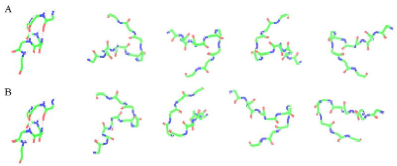
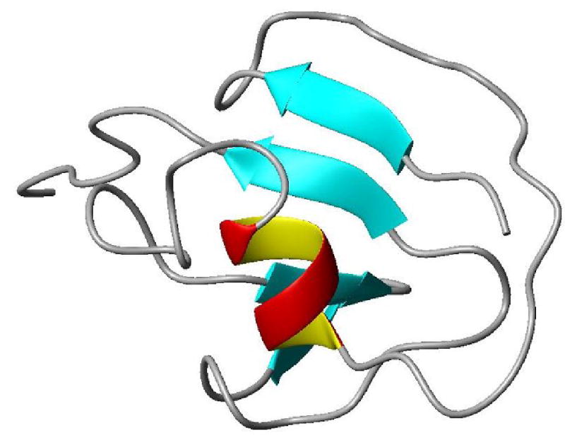
Structural genomics (or proteomics) activities are critically dependent on the availability of high-throughput structure determination methodology. Development of such methodology has been a particular challenge for NMR based structure determination because of the demands for isotopic labeling of proteins and the requirements for very long data acquisition times. We present here a methodology that gains efficiency from a focus on determination of backbone structures of proteins as opposed to full structures with all sidechains in place. This focus is appropriate given the presumption that many protein structures in the future will be built using computational methods that start from representative fold family structures and replace as many as 70% of the sidechains in the course of structure determination. The methodology we present is based primarily on residual dipolar couplings (RDCs), readily accessible NMR observables that constrain the orientation of backbone fragments irrespective of separation in space. A new software tool is described for the assembly of backbone fragments under RDC constraints and an application to a structural genomics target is presented. The target is an 8.7 kDa protein from Pyrococcus furiosus, PF1061, that was previously not well annotated, and had a nearest structurally characterized neighbor with only 33% sequence identity. The structure produced shows structural similarity to this sequence homologue, but also shows similarity to other proteins, which suggests a functional role in sulfur transfer. Given the backbone structure and a possible functional link this should be an ideal target for development of modeling methods.
Figures
Similar articles
-
Global folds of proteins with low densities of NOEs using residual dipolar couplings: application to the 370-residue maltodextrin-binding protein.J Mol Biol. 2000 Jun 30;300(1):197-212. doi: 10.1006/jmbi.2000.3842. J Mol Biol. 2000. PMID: 10864509
-
Determination of protein backbone structures from residual dipolar couplings.Methods Enzymol. 2005;394:175-209. doi: 10.1016/S0076-6879(05)94007-X. Methods Enzymol. 2005. PMID: 15808221 Free PMC article.
-
Structure determination of a new protein from backbone-centered NMR data and NMR-assisted structure prediction.Proteins. 2006 Nov 1;65(2):480-9. doi: 10.1002/prot.21119. Proteins. 2006. PMID: 16927360
-
Residual dipolar couplings: synergy between NMR and structural genomics.J Biomol NMR. 2002 Jan;22(1):1-8. doi: 10.1023/a:1013801714041. J Biomol NMR. 2002. PMID: 11885976 Review.
-
Incorporating residual dipolar couplings into the NMR solution structure determination of nucleic acids.Biopolymers. 1999-2000;52(4):168-80. doi: 10.1002/1097-0282(1999)52:4<168::AID-BIP1002>3.0.CO;2-7. Biopolymers. 1999. PMID: 11295749 Review.
Cited by
-
Simultaneous structure and dynamics of a membrane protein using REDCRAFT: membrane-bound form of Pf1 coat protein.J Magn Reson. 2010 Nov;207(1):8-16. doi: 10.1016/j.jmr.2010.07.016. Epub 2010 Jul 30. J Magn Reson. 2010. PMID: 20829084 Free PMC article.
-
Dynafold: a dynamic programming approach to protein backbone structure determination from minimal sets of Residual Dipolar Couplings.J Bioinform Comput Biol. 2014 Feb;12(1):1450002. doi: 10.1142/S0219720014500024. Epub 2014 Jan 7. J Bioinform Comput Biol. 2014. PMID: 24467760 Free PMC article.
-
REDCRAFT: a tool for simultaneous characterization of protein backbone structure and motion from RDC data.J Magn Reson. 2008 Apr;191(2):322-34. doi: 10.1016/j.jmr.2008.01.007. Epub 2008 Jan 16. J Magn Reson. 2008. PMID: 18258464 Free PMC article.
-
Structure-oriented methods for protein NMR data analysis.Prog Nucl Magn Reson Spectrosc. 2010 May;56(4):311-28. doi: 10.1016/j.pnmrs.2010.02.001. Epub 2010 Mar 3. Prog Nucl Magn Reson Spectrosc. 2010. PMID: 20633357 Free PMC article. Review. No abstract available.
-
Improvements to REDCRAFT: a software tool for simultaneous characterization of protein backbone structure and dynamics from residual dipolar couplings.J Biomol NMR. 2014 Dec;60(4):241-64. doi: 10.1007/s10858-014-9871-x. Epub 2014 Nov 18. J Biomol NMR. 2014. PMID: 25403759 Free PMC article.
References
-
- Montelione GT, Zheng DY, Huang YPJ, Gunsalus KC, Szyperski T. Protein NMR spectroscopy in structural genomics. Nature Structural Biology. 2000;7:982–985. - PubMed
-
- Tian F, Valafar H, Prestegard JH. A dipolar coupling based strategy for simultaneous resonance assignment and structure determination of protein backbones. J Am Chem Soc. 2001;123:11791–11796. - PubMed
-
- Orengo CA, Todd AE, Thornton JM. From protein structure to function. Curr Opin Struct Biol. 1999;9:374–382. - PubMed
-
- Sali A, Kuriyan J. Challenges at the frontiers of structural biology. Trends in Biochemical Sciences. 1999;24:M20–M24. - PubMed
-
- Holm L, Sander C. Database Algorithm for Generating Protein Backbone and Side-Chain Coordinates from a C-Alpha Trace Application to Model-Building and Detection of Coordinate Errors. J Mol Biol. 1991;218:183–194. - PubMed
Publication types
MeSH terms
Substances
Grants and funding
LinkOut - more resources
Full Text Sources
Other Literature Sources