Comparative Study
. 2005 Apr 15;564(Pt 2):437-50.
doi: 10.1113/jphysiol.2004.078725. Epub 2005 Feb 10.

Prostaglandin E2 potentiates a TTX-resistant sodium current in rat capsaicin-sensitive vagal pulmonary sensory neurones

Affiliations
Comparative Study

Prostaglandin E2 potentiates a TTX-resistant sodium current in rat capsaicin-sensitive vagal pulmonary sensory neurones

Kevin Kwong et al. J Physiol. .

Abstract

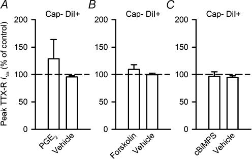
Capsaicin-sensitive vagal pulmonary neurones (pulmonary C neurones) play an important role in regulating airway function. During airway inflammation, the level of prostaglandin E(2) (PGE(2)) increases in the lungs and airways. PGE(2) has been shown to sensitize isolated pulmonary C neurones. The somatosensory correlate of the pulmonary C neurone, the small-diameter nociceptive neurone of the dorsal root ganglion, contains a high percentage of tetrodotoxin-resistant sodium currents (TTX-R I(Na)). Therefore, this study was carried out to determine whether these channel currents are involved in the PGE(2)-induced sensitization of pulmonary C neurones. We used the perforated patch-clamp technique to study the effects of PGE(2) on the TTX-R I(Na) in acutely cultured capsaicin-sensitive pulmonary neurones that were identified by retrograde labelling with a fluorescent tracer, DiI. We found that the pulmonary neurones sensitive to capsaicin had a higher percentage of TTX-R I(Na) than that of capsaicin-insensitive pulmonary neurones. PGE(2) exposure increased the evoked TTX-R I(Na) when experiments were performed at both room temperature and at 37 degrees C. Furthermore, stimulation of the adenylyl cyclase/protein kinase A pathway with either forskolin or Sp-5,6-DCl-cBiMPS potentiated the TTX-R I(Na) in a manner similar to that of PGE(2). We conclude that these modulatory effects of PGE(2) on TTX-R I(Na) play an important role in the sensitization of pulmonary C neurones.


Figures

Figure 1
Figure 1. A representative capsaicin-sensitive vagal pulmonary neurone illustrating that total inward INa is comprised of both TTX-S and TTX-R components
A, a family of total inward currents was generated from a voltage-clamp protocol in which 40 ms duration voltage steps from −70 to 45 mV in 5 mV increments (Vhold =−80 mV) were applied to a nodose ganglion pulmonary neurone (22.3 pF). Traces shown are the first 10 ms of each tracing of the 40 ms voltage pulse. B, the family of current tracings in the same neurone as A in the presence of 100 nm TTX. C, point-for-point subtraction of the TTX-R INa (B) from the total INa (A) yielded the TTX-S INa. D, current–voltage relationship showing differences in the peak current profiles among the INa species.
Figure 2
Figure 2. Capsaicin-sensitive vagal pulmonary neurones display a high percentage of TTX-R current
AJ, the TTX-R INa and total INa were obtained from the peak current of the family of evoked currents generated using the voltage step protocol described in Fig. 1. A, the subset of capsaicin-sensitive vagal pulmonary neurones showed a different percentage TTX-R INa–capacitance profile than that of capsaicin-insensitive vagal pulmonary neurones. B, comparison of percentage TTX-R INa between capsaicin-sensitive, DiI-labelled neurones (n = 28), which were presumed to be those that had innervated pulmonary structures, and capsaicin-insensitive pulmonary neurones (n = 23). Comparison of percentage TTX-R INa in the total populations of capsaicin-sensitive neurones (n = 43) and capsaicin-insensitive neurones (n = 37) in C, pulmonary neurones (n = 79) and non-pulmonary neurones (n = 17) in D, and jugular neurones (n = 38) and nodose neurones (n = 78) in E. F, a plot of the total INa as a function of cell capacitance. Comparisons of the total INa density between capsaicin-sensitive pulmonary neurones (n = 28) and capsaicin-insensitive pulmonary neurones (n = 23) in G, capsaicin-sensitive (n = 43) and capsaicin-insensitive neurones (n = 37) in H, pulmonary (n = 79) and non-pulmonary neurones (n = 17) in I, and jugular (n = 38) and nodose ganglion neurones (n = 78) in J. White circles, capsaicin-insensitive pulmonary neurones; grey circles, capsaicin-sensitive pulmonary neurones. Statistical comparisons were made using unpaired t tests. †P < 0.01; ‡P < 0.001.
Figure 3
Figure 3. PGE2 increases a voltage-dependent TTX-R INa
A, the representative experimental record depicts the rate at which the effects of PGE2 are manifested in a capsaicin-sensitive vagal pulmonary neurone (25.6 pF) from the nodose ganglion. The currents were evoked by a single 20 ms voltage step to −5 mV from Vhold (−80 mV; top). Raw tracings show an increase in the amplitude of evoked current during PGE2 perfusion (1 μm; bottom). The arrows indicate current traces recorded before (a) and after PGE2 treatment (b). For clarity, only the first 10 ms of the 20 ms voltage step tracings are shown. B, the plot of the peak TTX-R INa during PGE2 perfusion as a function of time is shown. The peak currents developed from a baseline level before PGE2 (a) to a maximal steady-state level during PGE2 perfusion (b) over the course of ∼ 5 min. Bar, duration of PGE2 perfusion. C, the experimental record was generated using the voltage-clamp protocol (top) to evoke a family of current tracings recorded before (middle) and after PGE2 treatment (1 μm; bottom) in a capsaicin-sensitive vagal pulmonary neurone (25.6 pF) harvested from the nodose ganglia. The full 40 ms voltage pulse was truncated for clarity. D, current density–voltage relationship showing that PGE2 pretreatment (1 μm; •) increased the TTX-R INa density over that of control (○) in capsaicin-sensitive vagal pulmonary neurones (n = 8).
Figure 4
Figure 4. PGE2 treatment potentiates the maximal conductance, voltage dependence and half-activation potential of the TTX-R INa in capsaicin-sensitive vagal pulmonary neurones
A, in capsaicin-sensitive vagal pulmonary neurones (n = 8), mean data show that treatment with PGE2 (1 μm; •) increased the maximal conductance compared with that of before treatment (○). The threshold of current activation was not different between experimental and control conditions. B, the conductance was normalized to the maximal conductance in order to better visualize the difference in voltage sensitivity and half-activation potential. PGE2 treatment (1 μm) appears to increase the voltage dependence and cause a hyperpolarizing shift in the half-activation potential compared with that of control. Each set of data was fitted to a single-term Boltzmann function.
Figure 5
Figure 5. Experimental record showing PGE2 modulation of steady state TTX-R INa in a capsaicin-sensitive vagal pulmonary neurone
A, the steady-state inactivation protocol consisted of holding the membrane at various prepulse potentials ranging from −90 mV to −5 mV for 500 ms and then stepping to a single depolarizing test potential of 10 mV for 20 ms (top). Raw traces from a nodose ganglion neurone (25.6 pF) document the changes recorded before (middle) and after PGE2 exposure (1 μm; bottom). B, group data showing that PGE2 increases steady-state TTX-R INa in capsaicin-sensitive vagal pulmonary neurones. Average data in capsaicin-sensitive vagal pulmonary neurones (n = 8) from nodose and jugular ganglia in which the steady-state TTX-R INa was plotted against the prepulse potential (top). The data illustrate that the TTX-R INa was dramatically potentiated after PGE2 treatment (•) compared with the currents evoked before treatment (○). In the expanded view of the steady-state inactivation curve that has been normalized to the maximal conductance (bottom), treatment with PGE2 (1 μm) appears to cause little increase in voltage dependence.
Figure 6
Figure 6. Group data illustrating the modulating effects of forskolin on capsaicin-sensitive vagal pulmonary neurones
A, mean data plotting the TTX-R INa density as a function of membrane voltage in capsaicin-sensitive vagal pulmonary neurones (n = 12). The plot compares the TTX-R INa density before (○) and after forskolin treatment (1 μm; •). B, forskolin increases maximal conductance of TTX-R INa in capsaicin-sensitive vagal pulmonary neurones. Group data plotting conductance as a function of membrane voltage before and after treating capsaicin-sensitive vagal pulmonary neurones with forskolin. Each set of data was fitted to a single-term Boltzmann function. C, forskolin increases half-activation potential and voltage dependence of TTX-R INa. Averaged data in the 12 capsaicin-sensitive vagal pulmonary neurones show that treatment with forskolin causes a steeper dependence on voltage compared with before treatment. Further, forskolin treatment shifted the half-activation potential in a hyperpolarized direction. Statistical analysis of A–C is represented in Table 2.
Figure 7
Figure 7. Modulation of TTX-R INa by forskolin or cBiMPS at room temperature (∼ 21°C) and 37°C
Currents were evoked by 20 ms steps to −5 mV from Vhold (−80 mV). A, raw tracings from the same neurone illustrating the modulation of evoked current during control and forskolin treatment (1 μm). B, mean data showing that peak evoked current at room temperature during forskolin treatment (n = 13) was greater than that during 1,9-dideoxyforskolin (10 μm; 1,9-ddf; P < 0.01; n = 6) and vehicle treatment (P < 0.01; n = 9). Statistical comparisons were evaluated using unpaired t tests. †Significantly different from forskolin treatment. C, peak evoked current recorded at 37°C was compared before and after a ∼ 10 min exposure to forskolin (1 μm) or vehicle. D, group data illustrating that at 37°C, forskolin significantly modulated TTX-R INa (n = 4) compared with that of vehicle treatment (P < 0.05; n = 4). Statistical comparisons were evaluated using unpaired t tests. *Significantly different from forskolin treatment. E, experimental record showing the potentiating effect of cBiMPS treatment on evoked TTX-R INa at 21°C compared with that of vehicle treatment. F, mean data (n = 6) showing that evoked currents were greater during cBiMPS (50 μm) than during vehicle treatment. Statistical comparisons were made using paired t tests. *Significantly different from cBiMPS treatment (P < 0.05).
Figure 8
Figure 8. Lack of modulation of TTX-R INa in capsaicin-insensitive pulmonary neurones
Currents evoked by 20 ms steps to −5 mV from Vhold (−80 mV). No difference (P > 0.05) was detected in evoked TTX-R INa during exposure to PGE2 (1 μm; n = 4) compared with vehicle (n = 3) in A, to forskolin (1 μm; n = 5) compared with vehicle (n = 3) in B, and to cBiMPS (50 μm; n = 6) compared with vehicle (n = 6) in C. Statistical comparisons were made using unpaired t tests.

Similar articles

Cited by

References

    1. Agostoni E, Chinnock JE, de Burgh Daly M, Murray JG. Functional and histological studies of the vagus nerve and its branches to the heart, lungs and abdominal viscera in the cat. J Physiol. 1957;135:182–205. - PMC - PubMed
    1. Akaike N. Gramicidin perforated patch recording and intracellular chloride activity in excitable cells. Prog Biophys Mol Biol. 1996;65:251–264. 10.1016/S0079-6107(96)00013-2. - DOI - PubMed
    1. Akaike N, Harata N. Nystatin perforated patch recording and its applications to analyses of intracellular mechanisms. Jpn J Physiol. 1994;44:433–473. 10.2170/jjphysiol.44.433. - DOI - PubMed
    1. Akins PT, McCleskey EW. Characterization of potassium currents in adult rat sensory neurons and modulation by opioids and cyclic AMP. Neuroscience. 1993;56:759–769. 10.1016/0306-4522(93)90372-M. - DOI - PubMed
    1. Akopian AN, Sivilotti L, Wood JN. A tetrodotoxin-resistant voltage-gated sodium channel expressed by sensory neurons. Nature. 1996;379:257–262. 10.1038/379257a0. - DOI - PubMed

Publication types

LinkOut - more resources