. 2005 Mar;17(3):804-21.
doi: 10.1105/tpc.104.030205. Epub 2005 Feb 10.

Light regulates COP1-mediated degradation of HFR1, a transcription factor essential for light signaling in Arabidopsis

Affiliations

Light regulates COP1-mediated degradation of HFR1, a transcription factor essential for light signaling in Arabidopsis

Jianping Yang et al. Plant Cell. 2005 Mar.

Abstract

Arabidopsis thaliana seedlings undergo photomorphogenesis in the light and etiolation in the dark. Long Hypocotyl in Far-Red 1 (HFR1), a basic helix-loop-helix transcription factor, is required for both phytochrome A-mediated far-red and cryptochrome 1-mediated blue light signaling. Here, we report that HFR1 is a short-lived protein in darkness and is degraded through a 26S proteasome-dependent pathway. Light, irrespective of its quality, enhances HFR1 protein accumulation via promoting its stabilization. We demonstrate that HFR1 physically interacts with Constitutive Photomorphogenesis 1 (COP1) and that COP1 exhibits ubiquitin ligase activity toward HFR1 in vitro. In addition, we show that COP1 is required for degradation of HFR1 in vivo. Furthermore, plants overexpressing a C-terminal 161-amino acid fragment of HFR1 (CT161) display enhanced photomorphogenesis, suggesting an autonomous function of CT161 in promoting light signaling. This truncated HFR1 gene product is more stable than the full-length HFR1 protein in darkness, indicating that the COP1-interacting N-terminal portion of HFR1 is essential for COP1-mediated destabilization of HFR1. These results suggest that light enhances HFR1 protein accumulation by abrogating COP1-mediated degradation of HFR1, which is necessary and sufficient for promoting light signaling. Additionally, our results substantiate the E3 ligase activity of COP1 and its critical role in desensitizing light signaling.


Figures

Figure 1.
Figure 1.
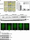
Hypersensitive Light Responses of HFR1 Overexpression Lines. (A) Morphology of Arabidopsis seedlings overexpressing Myc-HFR1 or GFP-HFR1 grown under various light conditions. Dk, darkness; FR, far-red; R, red; B, blue light. Photographs of seedlings from each light condition were taken at the same magnification. Bars = 1 mm. (B) Quantification of hypocotyl lengths (average of 20 seedlings) under various light conditions. Bars stand for standard deviations. (C) Light-responsive gene expression (CAB3, RBCS, and CHS). Seedlings were grown in darkness (Dk) for 4 d then transferred into far-red light (FR) for 12 h. An rRNA UV fluorescence image of the duplicating gels was shown as a loading control. Transgenic line A2 and transgenic line D3 were used as the representative lines for Myc-HFR1 and GFP-HFR1, respectively, unless otherwise indicated. (D) RNA gel blot analysis of Myc-HFR1 and GFP-HFR1 transgene expression. Seedlings were grown under continuous far-red light for 5 d. An rRNA UV fluorescence image of the gel was shown as a loading control.
Figure 2.
Figure 2.
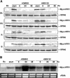
Light Enhances HFR1 Protein Accumulation. (A) Morphology of 4-d-old Myc-HFR1 (top) and GFP-HFR1 (bottom) transgenic seedlings grown under various fluence rates of white light. Seedlings shown in the left panels were grown under low light intensity (∼10 μmol/m2s), seedlings shown in the middle panels were grown under medium light intensity (∼100 μmol/m2s), and the seedlings shown in the right panels were grown under high light intensity (∼500 μmol/m2s). Bars = 1 mm. (B) Quantification of hypocotyl lengths of the seedlings grown under various fluence rates of white light. Bars stand for standard deviations. (C) Immunoblot analyses of Myc-HFR1 in seedlings grown in continuous darkness (Dk) for 4 d and then transferred to white light of different fluence rates for the indicated amounts of time. An anti-RPN6 (a 26S proteasome subunit) immunoblot is shown below to indicate approximately equal loadings. (D) Enhanced GFP-HFR1 fluorescence and nuclear accumulation (confirmed by 4′,6-diamidino-2-phenylindole staining; data not shown) in hypocotyls cells under white light (WL) for 4 h or far-red (FR), red (R), and blue (B) light for 6 h. Dk, dark-grown seedlings. Photographs of seedlings were taken at the same magnification. Bar = 50 μm. (E) Various colors of light enhance HFR1 accumulation during dark-to-light transition. Arabidopsis seedlings expressing Myc-HFR1 or GFP-HFR1 were grown in darkness (Dk) for 4 d and then transferred to various light conditions for 6 h before total protein was extracted for immunoblot analysis. Immunoblots of anti-RPN6 were shown below to indicate approximately equal loadings. FR, far-red; R, red; B, blue; WL, white light.
Figure 3.
Figure 3.
HFR1 Protein Is Rapidly Degraded through a Proteasome-Dependent Pathway. (A) and (B) Myc-HFR1 and GFP-HFR1 proteins are stabilized by the proteasome inhibitor MG132. Arabidopsis seedlings expressing Myc-HFR1 or GFP-HFR1 were grown in darkness for 4 d and then treated with MG132 or mock treated with 0.1% DMSO for 6 h. Total protein was extracted and subjected to immunoblot analysis. (C) Fluorescence images of hypocotyl cells of Arabidopsis seedlings expressing GFP-HFR1. Seedlings were grown in darkness for 4 d and then treated with MG132 or mock treated with 0.1% DMSO for 24 h. Photographs of seedlings were taken at the same magnification. Bar = 50 μm. (D) Arabidopsis seedlings expressing Myc-HFR1 or GFP-HFR1 were grown in darkness for 4 d and then treated with MG132 for 24 h to promote accumulation of the fusion proteins. The seedlings were then washed thoroughly and incubated in darkness or white light (WL) for indicated amounts of time. Total protein was extracted and subjected to immunoblot analysis. Immunoblots of anti-RPN6 were shown below to indicate approximately equal loadings.
Figure 4.
Figure 4.
Light-Quality Control of HFR1 Protein Accumulation. (A) Differential effects of light qualities on HFR1 protein accumulation. Arabidopsis seedlings expressing Myc-HFR1 were grown in continuous red light (Rc) for 4 d and then treated with MG132 for 24 h to promote the accumulation of HFR1 protein (start point). The seedlings were then incubated in darkness in the presence of DMSO or MG132 for the indicated amounts of time before harvested for immunoblot analyses. Immunoblots of anti-RPN6 were shown below to indicate approximately equal loadings. (B) Effects of DMSO and MG132 on Myc-HFR1 transcript accumulation. Arabidopsis seedlings were grown in continuous red light (Rc) for 4 d and then treated with MG132 for 24 h. The seedlings were then transferred to darkness for the indicated times in the presence of DMSO or MG132 before total RNAs were extracted for RNA gel blot analysis. A UV fluorescence image of rRNA of the gel is shown as a loading control.
Figure 5.
Figure 5.
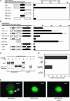
HFR1 Physically Interacts with COP1. (A) and (B) HFR1–COP1 interaction analyzed by the yeast two-hybrid assay. Left panels illustrate the prey and bait constructs, and the right panels show the corresponding β-galactosidase activities. The value is the average of six individual yeast colonies, and the error bars represent the standard deviations. (C) In vitro coimmunoprecipitation of COP1 by HFR1. The 35S-labeled COP1 or COP1-ΔCC were incubated with partially labeled GAD-HFR1 or GAD and coimmunoprecipitated with anti-GAD antibodies. Supernatant fractions (1.6%) and pellet fractions (33.3%) were resolved by SDS-PAGE and visualized by autoradiography using a phosphor imager. Successful immunoprecipitation of GAD was confirmed on a separate gel (data not shown). (D) Quantification of the fractions of bound prey proteins. Error bars denote one standard error of the mean of two replicate experiments. (E) Recruitment of GFP-NT131 into COP1 nuclear speckles in living onion epidermal cells. The left panel shows that GFP-COP1 is localized in bright nuclear speckles and the fluorescent cytoplasmic inclusion body (IB; indicated by an arrow). The middle panel shows that GFP-NT131 is uniformly distributed in the nucleus. Coexpression of GFP-NT131 with nontagged COP1 resulted in bright green nuclear speckles over the uniform green fluorescent background. This result suggests a direct interaction between COP1 and HFR1 in living plant cells. Dashed lines demarcate the nuclei (confirmed by 4′,6-diamidino-2-phenylindole staining; data not shown). Bars = 50 μm.
Figure 6.
Figure 6.
In Vitro Ubiquitination Assay of HFR1. GST-tagged NT131 of HFR1 (GST-NT131) protein was used as the substrate. (A) An anti-HFR1 immunoblot showing ubiquitinated GST-NT131 (indicated by an asterisk). (B) An immunoblot showing ubiquitinated HFR1 detected by streptavidin-conjugated horseradish peroxidase for biotinylated ubiquitin, followed by chemiluminescence visualization. The arrowhead indicates unmodified GST-NT131.
Figure 7.
Figure 7.
HFR1 Protein Degradation Is Defective in cop1 Mutants. (A) Seedling morphology showing that the cop1 mutant phenotype is enhanced by the GFP-HFR1 transgene under all light conditions. Photographs of seedlings from each light condition except darkness were taken at the same magnification. For dark-grown seedlings, the three seedlings on the left side were of the same magnification, whereas the four seedlings on the right side were of another magnification. Dk, darkness; FR, far red; R, red; B, blue. Bars = 1 mm. (B) Quantification of hypocotyl lengths of various mutants under different light conditions. Bars stand for standard deviations. (C) Fluorescence images of GFP-HFR1 in dark-grown root cells. Panel 1, parental GFP-HFR1 transgenic seedlings, line B4; panels 2 and 3, cop1-4 and cop1-6 seedlings harboring the GFP-HFR1 transgene, respectively. Bar = 50 μm. (D) Effects of cop1 mutations on HFR1 protein accumulation in seedlings grown either in continuous darkness for 5 d or grown in darkness for 5 d then transferred to white light for 1 h. An immunoblot of anti-RPN6 is shown below to indicate approximately equal loadings. Lane 1, parental GFP-HFR1 transgenic seedlings, line B4; lanes 2 and 3, cop1-4 and cop1-6 seedlings, respectively, harboring the GFP-HFR1 transgene. (E) Effects of cop1 mutations on HFR1 transcript accumulation under different light conditions. Seedlings were grown in darkness for 5 d or grown in darkness (D) for 4 d and then transferred to continuous far-red (FR), red (R), or blue light (B) for 24 h. In each blot, HFR1 transcript is shown on the top and a UV fluorescence image of the rRNA is shown below as a loading control.
Figure 8.
Figure 8.
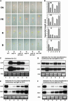
Epistasis Analyses of hfr1-201 and cop1 Mutations. (A) Seedling morphology of seedlings grown under continuous darkness (Dk) and far-red (FR), red (R), and blue (B) light conditions. Photographs of seedlings from each light condition were taken at the same magnification. The genotypes of seedlings shown in all panels in this figure are as follows: 1, the wild type; 2, cop1-4; 3, cop1-4 hfr1-201; 4, cop1-6; 5, cop1-6 hfr1-201; 6, hfr1-201. All mutations are in the same ecotype background (Columbia). Bars = 1 mm. (B) Quantification of hypocotyl lengths. Bars stand for standard deviations. (C) CAB3 and RBCS expression in dark-grown seedlings. (D) CAB3 and RBCS expression seedlings grown in darkness for 4 d and then transferred to far-red light for 24 h. (E) CAB3 and RBCS expression seedlings grown in darkness for 4 d and then transferred to red light for 24 h. (F) CAB3 and RBCS expression seedlings grown in darkness for 4 d and then transferred to blue light for 24 h. For (C) to (F), a representative UV fluorescence image of rRNA from the duplicating gels of each light condition is shown as a loading control.
Figure 9.
Figure 9.
CT161 of HFR1 Is Functional Autonomous in Promoting Light Signaling, whereas NT131 Is Required for COP1-Mediated Destabilization of HFR1. (A) Overexpression GFP-CT161 in the hfr1-201 mutant background causes a strong hypersensitive response to far-red (FR), red (R), and blue light (B) and normal etiolation growth in the dark (Dk). Two independent lines are shown. Photographs of seedlings from each light condition were taken at the same magnification. WT, wild-type plants. Bars = 1mm. (B) Quantification of hypocotyl lengths under various light conditions. Bars stand for standard deviations. (C) Immunoblot showing that GFP-CT161 accumulates in dark-grown seedlings, whereas no GFP-HFR1 was detected in the absence of MG132. MG132 treatment stabilizes GFP-HFR1 but has minimal effects on the accumulation of GFP-CT161. An immunoblot of anti-RPN6 was shown below to indicate approximately equal loadings. Lane 1, GFP-HFR1 transgenic line D3; lanes 2 and 3, GFP-CT161 transgenic lines B3 and D1, respectively. (D) Effects of hfr1-201 mutation, GFP-HFR1, and GFP-CT161 transgenes on HY5 accumulation. Seedlings were grown in continuous darkness, far-red, red, or blue light for 5 d, then total protein was extracted for immunoblot analysis. Immunoblots of anti-RPN6 are shown below to indicate approximately equal loadings. Lane 1, the wild type; lane 2, hfr1-201 mutants; lane 3, GFP-HFR1 seedlings; lane 4, GFP-CT161 seedlings.
Figure 10.
Figure 10.
A Model Depicting the Structure–Function Relationship of HFR1. The N-terminal 131–amino acid fragment (NT131) is involved in interaction with COP1 and is essential for COP1-mediated destabilization of HFR1. The C-terminal 161–amino acid fragment (CT161) is autonomous in DNA binding and promoting photomorphogenesis. Light abrogates COP1-mediated destabilization of HFR1 by depleting the nuclear abundance of COP1 (represented by a bar). ++, putative nuclear localization signals.

Similar articles

Cited by

References

    1. Ahmad, M., Jarillo, J.A., Smirnova, O., and Cashmore, A.R. (1998). The CRY1 blue light photoreceptor of Arabidopsis interacts with phytochrome A in vitro. Mol. Cell 1, 939–948. - PubMed
    1. Ang, L.-H., Chattopadhyay, S., Wei, N., Oyama, T., Okada, K., Batschauer, A., and Deng, X.W. (1998). Molecular interaction between COP1 and HY5 defines a regulatory switch for light control of Arabidopsis development. Mol. Cell 1, 213–222. - PubMed
    1. Bauer, D., Viczián, A., Kircher, S., Nobis, T., Nitschke, R., Kunkel, T., Panigrahi, K.C.S., Ádám, E., Fejes, E., Schäfer, E., and Nagy, F. (2004). Constitutive photomorphogenesis 1 and multiple photoreceptors control degradation of phytochrome interacting factor 3, a transcription factor required for light signaling in Arabidopsis. Plant Cell 16, 1433–1445. - PMC - PubMed
    1. Benvenuto, G., Formiggini, F., Laflamme, P., Malakhov, M., and Bowler, C. (2002). The photomorphogenesis regulator DET1 binds the amino-terminal tail of histone H2B in a nucleosome context. Curr. Biol. 12, 1529–1534. - PubMed
    1. Briggs, W.R., and Olney, M.A. (2001). Photoreceptors in plant photomorphogenesis to date: Five phytochromes, two cryptochromes, one phototropin, and one superchrome. Plant Physiol. 125, 85–88. - PMC - PubMed

Publication types

MeSH terms