. 2005 Mar;17(3):876-87.
doi: 10.1105/tpc.104.026872. Epub 2005 Feb 10.

A vacuolar processing enzyme, deltaVPE, is involved in seed coat formation at the early stage of seed development

Affiliations

A vacuolar processing enzyme, deltaVPE, is involved in seed coat formation at the early stage of seed development

Satoru Nakaune et al. Plant Cell. 2005 Mar.

Abstract

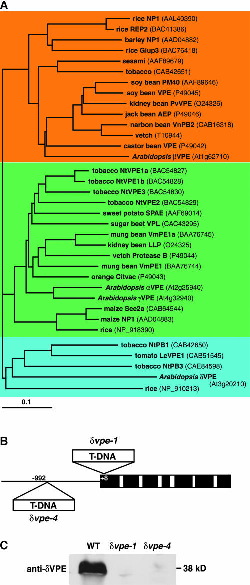
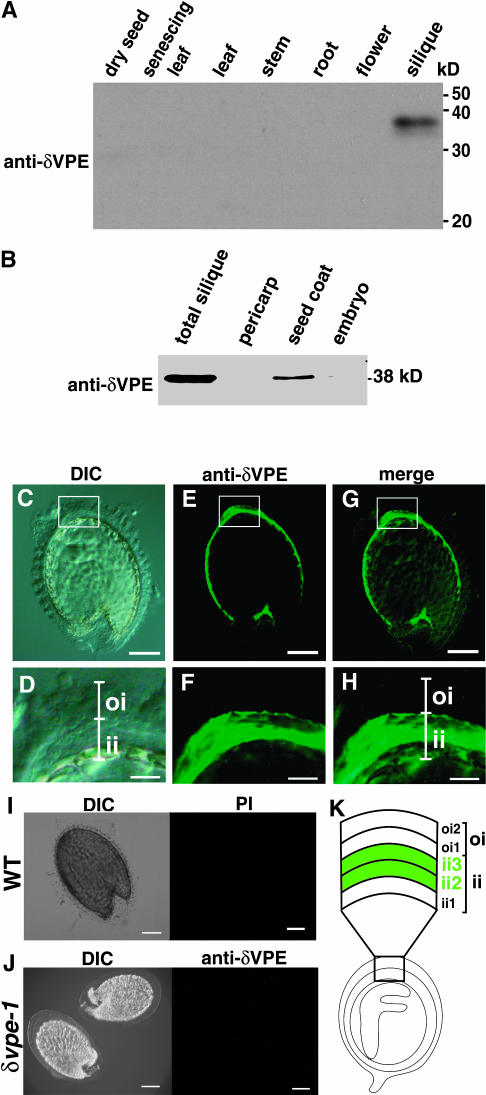
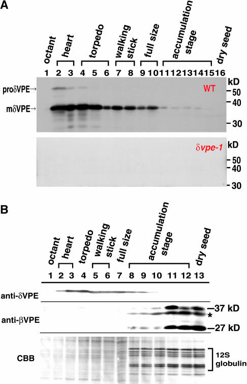
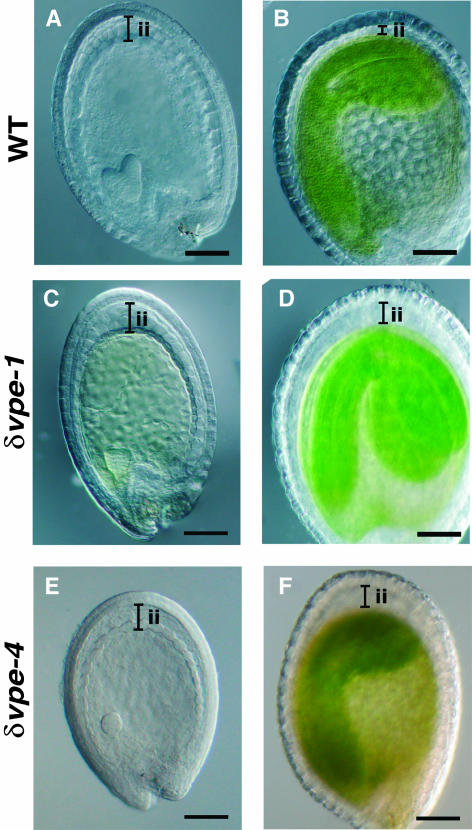
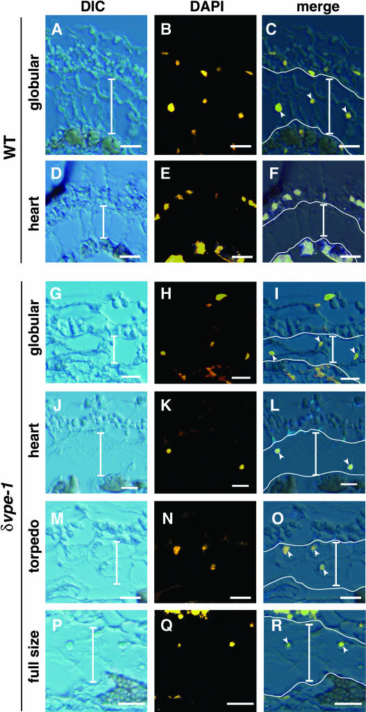
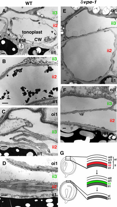
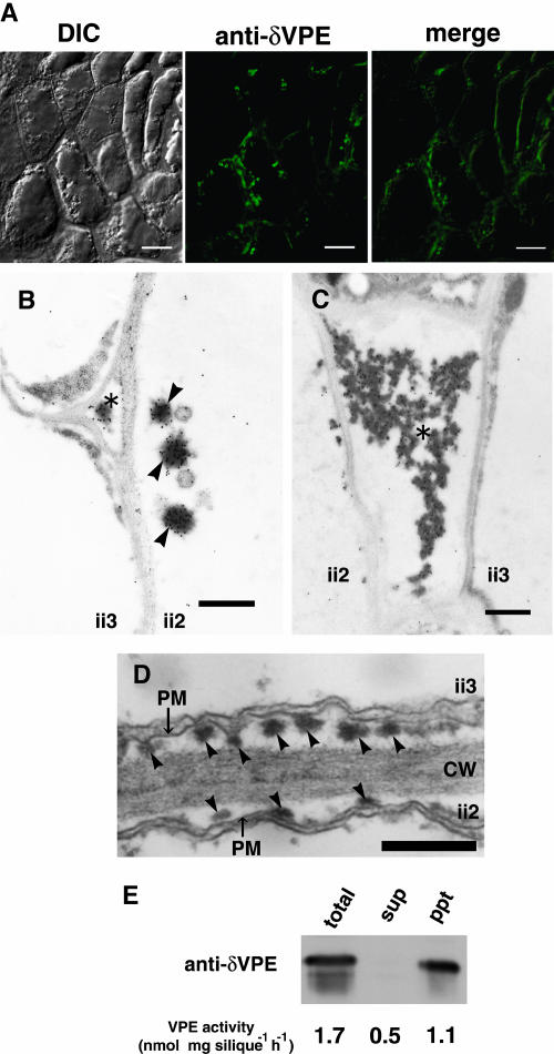
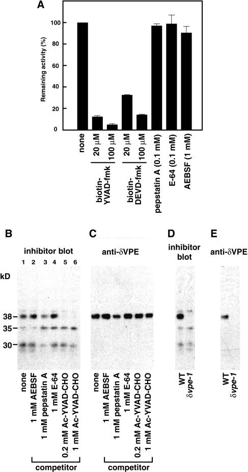
Vacuolar processing enzyme (VPE) is a Cys proteinase responsible for the maturation of vacuolar proteins. Arabidopsis thaliana deltaVPE, which was recently found in the database, was specifically and transiently expressed in two cell layers of the seed coat (ii2 and ii3) at an early stage of seed development. At this stage, cell death accompanying cell shrinkage occurs in the ii2 layer followed by cell death in the ii3 layer. In a deltaVPE-deficient mutant, cell death of the two layers of the seed coat was delayed. Immunocytochemical analysis localized deltaVPE to electron-dense structures inside and outside the walls of seed coat cells that undergo cell death. Interestingly, deltaVPE in the precipitate fraction from young siliques exhibits caspase-1-like activity, which has been detected in various types of plant cell death. Our results suggest that, at the early stage of seed development, deltaVPE is involved in cell death of limited cell layers, the purpose of which is to form a seed coat.


Figures

Figure 1.
Figure 1.
Plant VPEs and Arabidopsis δVPE Knockout Mutants. (A) A dendrogram of plant VPEs. VPEs are separated into three types: a vegetative type (green), an embryo type (orange), and a novel type (blue). The dendrogram was drawn with the ClustalW and TreeView programs. The horizontal scale represents the evolutionary distance expressed as the number of substitutions per amino acid. (B) Schematic representation of the Arabidopsis δVPE gene and the positions of the T-DNA insertions in the δvpe-1 and δvpe-4 alleles. Black boxes indicate exons, open boxes indicate introns, and a solid line indicates the 5′-nonconding region. (C) Immunoblot analysis of a young silique from the wild-type and δvpe knockout mutants (δvpe-1 and δvpe-4) with antibodies directed against δVPE.
Figure 2.
Figure 2.
δVPE Is Specifically Localized in Two Cell Layers of the Inner Integuments of the Seed Coats in Developing Arabidopsis Seeds. (A) Immunoblot analysis showing the organ-specific expression of δVPE. Total proteins were extracted from each organ (2 mg fresh weight) of Arabidopsis plants and were subjected to immunoblotting with anti-δVPE antibodies. The molecular masses are given on the right in kilodaltons. (B) Immunoblot analysis showing the tissue-specific expression of δVPE. Total proteins were extracted from each tissue of one young silique and were subjected to immunoblotting with anti-δVPE antibodies. (C) to (H) Immunofluorescence analysis of developing seeds at walking-stick-shaped-embryo stage with anti-δVPE antibodies. Green fluorescent images (anti-δVPE), differential interference contrast (DIC) images, and merged images (merge) are shown. (D), (F), and (H) are enlarged images of boxed regions in (C), (E), and (G), respectively. oi, outer integument; ii, inner integument. Bars = 100 μm in (C), (E), and (G) and 25 μm in (D), (F), and (H). (I) A control experiment with developing wild-type seeds and preimmune serum (PI). Bars = 100 μm. (J) A control experiment with developing δvpe-1 seeds and anti-δVPE antibodies. Bars = 100 μm. (K) An illustration of the organization of the cell layers in the seed coat of a developing seed having a walking-stick-shaped embryo. The seed coat is composed of two kinds of integuments: outer integument (oi) of two cell layers (oi1 and oi2) and inner integument (ii) of three cell layers (ii1, ii2, and ii3). Green indicates the cell layers (ii2 and ii3) that express δVPE.
Figure 3.
Figure 3.
δVPE Is Expressed at an Early Stage in Developing Arabidopsis Seeds, whereas βVPE Is Expressed in the Late Stage in Association with the Accumulation of Seed Storage Proteins. (A) Developmental changes in the levels of the precursor and mature forms of δVPE during seed development. The wild-type and δvpe-1 siliques were harvested at various stages, and one-tenth of total proteins from developing seeds in each one silique were subjected to immunoblotting with anti-δVPE antibodies. Developmental stages are indicated by shapes of embryo in the seeds: octant, four- to eight-celled embryo (lane 1); heart, heart-shaped embryo (lanes 2 and 3); torpedo, torpedo-shaped embryo (lanes 4 to 6); walking stick, walking-stick-shaped embryo (lanes 7 and 8), full size, almost full-sized embryo (lanes 9 and 10), accumulation stage, embryo accumulating seed storage proteins (lanes 11 To 15); dry seed (lane 16). The molecular masses are given at the right in kilodaltons. (B) Comparison of the expression pattern of δVPE with that of βVPE during seed development. The wild-type siliques were harvested at the stages indicated in (A), and one-tenth of total proteins from developing wild-type seeds in each silique was subjected to immunoblotting with anti-δVPE antibodies (anti-δVPE) or anti-βVPE antibodies (anti-βVPE) and to Coomassie blue staining (CBB). The 27- and 37-kD bands are two forms of βVPE. 12S globulin is a major storage protein. Asterisk indicates a nonspecific signal.
Figure 4.
Figure 4.
Thickness of Inner Integuments of the Seed Coats Is Reduced in the Wild-Type Seeds at the Early Stages, whereas It Is Not Reduced in the δvpe Seeds. Differential interference contrast images of developing seeds at an early-embryo stage ([A], [C], and [E]) and walking-stick-shaped-embryo stage ([B], [D], and [F]) from the wild type ([A] and [B]), δvpe-1 ([C] and [D]), and δvpe-4 ([E] and [F]). ii, inner integument. Bars = 100 μm.
Figure 5.
Figure 5.
Degradation of Nuclei in ii2 and ii3 Cell Layers of the Inner Integuments Occurs in the Wild-Type Seeds at the Heart-Shaped-Embryo Stage, whereas It Does Not Occur in the δvpe-1 Seeds at the Early Stages. Differential interference contrast (DIC) images, DAPI images, and merged images (merge) of developing seeds from the wild type ([A] to [F]) and δvpe-1 ([G] to [R]) are shown. DAPI images were visualized using 365/12-nm-wavelength excitation and a long-pass 397-nm-wavelength emission filter and then were converted in yellow to highlight the nuclei. Developmental stages are indicated at the left. Vertical bars indicate both ii2 and ii3 cell layers. Arrowheads indicate nuclei stainable with DAPI in the ii2 and ii3 cell layers. The cells of ii2 and ii3 layers, which are highly vacuolated, are larger than the cells of the other layers. The cells of outer integument layers contain a lot of starch granules, and the cells of ii1 layer accumulate pigments, which generated autofluorescence. Horizontal bars = 25 μm.
Figure 6.
Figure 6.
Morphological Changes of ii2 and ii3 Cell Layers of the Inner Integuments in the Developing Wild-Type and δvpe-1 Seeds at the Torpedo-Shaped-Embryo Stage. (A) Cell shrinkage and plasmolysis occurs first in the ii2 cell layers of developing wild-type seeds. Plasma membrane and tonoplast are partially disrupted (asterisk). At this stage, cells in the ii1 and ii3 layers are intact. Electron-dense structures (arrowheads) appear in the space between the plasma membrane (PM) and cell wall (CW). (B) The plasma membranes and cellular organelles are disrupted in cells of the ii2 layers in developing wild-type seeds. (C) and (D) Dead cells of the ii2 layers in developing wild-type seeds are finally compressed. (E) and (F) Cells of the ii2 and ii3 layers in developing δvpe-1 seeds remain intact. (G) An illustration of the disappearance of the ii2 cell layer (red) followed by that of the ii3 cell layers (green) in developing wild-type seeds. Bars = 1 μm (A) to (F).
Figure 7.
Figure 7.
Localization of δVPE in Electron-Dense Structures of Shrinking Cells in the Inner Integuments of Developing Seeds at the Torpedo-Shaped-Embryo Stage. (A) Immunofluorescence analysis of surfaces of the inner integuments with anti-δVPE antibodies. A green fluorescent image (anti-δVPE), a differential interference contrast (DIC) image, and a merged image (merge) are shown. Bars = 10 μm. (B) and (C) Immunoelectron micrographs of the inner integuments with anti-δVPE antibodies showing the localization of δVPE in electron-dense structures outside (asterisks) and inside (arrowheads) cell walls. Bars = 500 nm. (D) Electron micrograph showing the existence of electron-dense structures (arrowheads) in the space between the plasma membrane (PM) and cell wall (CW) of developing seeds of δvpe-1. Bars = 500 nm. (E) Accumulation of δVPE protein and VPE activity in the pellet fraction of young siliques. Total protein from young siliques was centrifuged to obtain the supernatant (sup) and precipitate (ppt) fractions. Each fraction was subjected to immunoblot analysis with anti-δVPE antibodies and to an assay of VPE activity.
Figure 8.
Figure 8.
δVPE Exhibits Caspase-1–Like Activity. (A) Effects of various proteinase inhibitors on the activity of VPE. Extracts from the developing seeds at the torpedo-shaped-embryo stage were preincubated with each proteinase inhibitor (as indicated) and then subjected to a VPE assay. (B) and (C) Extracts from the developing seeds at the torpedo-shaped-embryo stage were preincubated with each proteinase inhibitor (as indicated) before incubation with biotin-YVAD-fmk and then subjected to a biotinylated inhibitor blot (B) and to an immunoblot with anti-δVPE antibodies (C). Proteinase inhibitors used as a competitor: AEBSF, Ser protease inhibitor; pepstatin A, aspartic protease inhibitor; E-64, papain-type Cys protease inhibitor; Ac-YVAD-CHO, caspase-1 inhibitor. (D) and (E) Extracts from developing seeds of the wild type and δvpe-1 were subjected to a biotinylated inhibitor blot (D) and to an immunoblot with anti-δVPE antibodies (E).

Similar articles

Cited by

References

    1. Asamizu, E., Nakamura, Y., Sato, S., and Tabata, S. (2000). A large scale analysis of cDNA in Arabidopsis thaliana: Generation of 12,028 non-redundant expressed sequence tags from normalized and size-selected cDNA libraries. DNA Res. 7, 175–180. - PubMed
    1. Becker, C., Shutov, A.D., Nong, V.H., Senyuk, V.I., Jung, R., Horstmann, C., Fischer, J., Nielsen, N.C., and Muntz, K. (1995). Purification, cDNA cloning and characterization of proteinase B, an asparagine-specific endopeptidase from germinating vetch (Vicia sativa L.) seeds. Eur. J. Biochem. 228, 456–462. - PubMed
    1. Beeckman, T., De Rycke, R., Viane, R., and Inze, D. (2000). Histological study of seed coat development in Arabidopsis thaliana. J. Plant Res. 113, 139–148.
    1. Caffrey, C.R., Mathieu, M.A., Gaffney, A.M., Salter, J.P., Sajid, M., Lucas, K.D., Franklin, C., Bogyo, M., and McKerrow, J.H. (2000). Identification of a cDNA encoding an active asparaginyl endopeptidase of Schistosoma mansoni and its expression in Pichia pastoris. FEBS Lett. 466, 244–248. - PubMed
    1. Chen, J.M., Dando, P.M., Stevens, R.A., Fortunato, M., and Barrett, A.J. (1998). Cloning and expression of mouse legumain, a lysosomal endopeptidase. Biochem. J. 338, 111–117. - PMC - PubMed

Publication types

MeSH terms