. 2004 Jul-Dec;10(7-12):104-11.
doi: 10.2119/2004-00027.Tayebi.

Disease-associated prion protein elicits immunoglobulin M responses in vivo

Affiliations

Disease-associated prion protein elicits immunoglobulin M responses in vivo

Mourad Tayebi et al. Mol Med. 2004 Jul-Dec.

Abstract

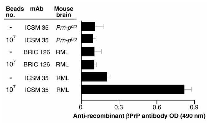
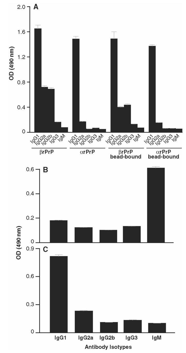
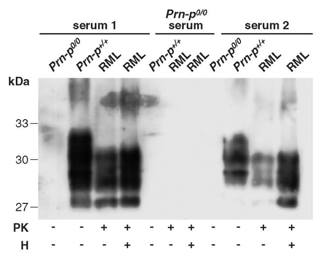
Prion diseases such as Creutzfeldt-Jakob disease are believed to result from the misfolding of a widely expressed normal cellular prion protein, PrPc. The resulting disease-associated isoforms, PrP(Sc), have much higher beta-sheet content, are insoluble in detergents, and acquire relative resistance to proteases. Although known to be highly aggregated and to form amyloid fibrils, the molecular architecture of PrP9Sc) is poorly understood. To date, it has been impossible to elicit antibodies to native PrP(Sc) that are capable of recognizing PrP(Sc) without denaturation, even in Pm-P(o/o) mice that are intolerant of it. Here we demonstrate that antibodies for native PrPc and PrP(Sc) can be produced by immunization of Pm-P(o/o) mice with partially purified PrPc and PrP(Sc) adsorbed to immunomagnetic particles using high-affinity anti-PrP monoclonal antibodies (mAbs). Interestingly, the polyclonal response to PrP(Sc) was predominantly of the immunoglobulin M (IgM) isotype, unlike the immunoglobulin G (IgG) responses elicited by PrP(c) or by recombinant PrP adsorbed or not to immunomagnetic particles, presumably reflecting the polymeric structure of disease-associated prion protein. Although heat-denatured PrP(Sc) elicited more diverse antibodies with the revelation of C-terminal epitopes, remarkably, these were also predominantly IgM suggesting that the increasing immunogenicity, acquisition of protease sensitivity, and reduction in infectivity induced by heat are not associated with dissociation of the PrP molecules in the diseased-associated protein. Adsorbing native proteins to immunomagnetic particles may have general applicability for raising polyclonal or monoclonal antibodies to any native protein, without attempting laborious purification steps that might affect protein conformation.


Figures

Figure 1
Figure 1
Adsorption of PrPSc is proportional to ICSM 35 concentration. Decreasing 10-fold dilutions of ICSM 35 (starting at 50 mg/mL) were adsorbed to Dynabeads as described in Materials and Methods. Next 107 of these beads were added to 50 μL of a 10% RML brain homogenate spiked into an equal volume of 10% Prn-Pº/º brain homogenate. Representative of 3 experiments.
Figure 2
Figure 2
Optimal detection and depletion of PrPSc from homogenized brain. RML brain homogenate spiked into brain homogenate derived from Prn-Pº/º mice was proteinase K (PK)-treated then incubated with 107 ICSM 35 saturated Dynabeads (upper panel). In the lower panel, ICSM 35 was omitted from the immunocomplex, keeping the other conditions identical. Representative of 3 experiments.
Figure 3
Figure 3
Polyclonal antibody responses to bead-adsorbed prion proteins including normal (•) and RML (▴) brain homogenates, and rPrP (▪) in FVB-N Prn-Pº/º mice (n = 3). Several specificity controls were used including omitting the homogenate/antigen (♦), adsorbing antigens from Prn-Pº/º mice (▿), or using an irrelevant isotype-matched mAb control for adsorption.
Figure 5
Figure 5
Pep-scan of sera generated with bead-bound prion proteins. A: PrPSc-Dynabeads sera from Prn-Pº/º mice were assayed against 20-mer peptides spanning the 91–231 region of the mouse PrP protein. B: PrPSc-Dynabeads (heat-treated prior to adsorption) sera from Prn-Pº/º mice were assayed against 20-mer peptides spanning the 91–231 region of the mouse PrP protein. C: PrPc-Dynabeads sera from Prn-Pº/º mice were assayed against 20-mer peptides spanning the 91–231 region of the mouse PrP protein.
Figure 4
Figure 4
Polyclonal antibodies from mice immunized with PrPSc-Dynabeads recognize mouse rβPrP in ELISA. Anti-PrP responses were assayed in mice (n = 5 for each arm) immunized with RML (PrPSc) or Prn-Pº/º brain homogenates adsorbed or not to antibody (ICSM or an isotype control mAb BRIC 126)-coated sheep anti-mouse Dynabeads-M450. PrP was adsorbed from 100 μL of a 1% PK-digested homogenate, or for arms without Dynabeads, 100 μL of a 1% homogenate emulsified in Freunds was used for immunization. Sheep anti-mouse Dynabeads-M450 coated with anti-PrP (ICSM 35) or an isotype-match control mAb (BRIC 126) were used to adsorb after proteinase K digestion. After omission of the Dynabeads, anti-PrP (ICSM 35) or isotype-match control mAb (BRIC 126) were used to bind RML (PrPSc) or Prn-Pº/º brain homogenates. Serum were assayed at 1:100 dilution. The assay negative control (Prn-Pº/º mouse serum) gave optical density (OD) values of 0.1 ± 0.03.
Figure 6
Figure 6
Isotype profile of anti-PrP polyclonal responses. FVB-N Prn-Pº/º mice were immunized with either soluble or bead-bound recombinant α or β PrP91–231 (panel A), PrPSc-Dynabeads (panel B), or PrPc-Dynabeads (panel C), and the isotype of anti-PrP produced was determined using Ig subclass–specific secondary antibodies in the standard recombinant PrP ELISA (see Materials and Methods). Data points represent the mean serum OD values of 1.24 ± 0.41, derived from 5 mice immunized with each antigen. Sera from unimmunized mice or mice similarly immunized with CFA/IFA emulsified with PBS gave OD values < 0.05. Representative of 2 experiments.
Figure 7
Figure 7
Immunoprecipitation of mouse PrP using polyclonal sera derived from Prn-Pº/º mice immunized (serum 1 or 2) or not (Prn-Pº/º) with PrPSc-Dynabeads. These sera were incubated with brain homogenates prepared from scrapie-affected and normal animals. Bound antibody was immuno-precipitated with protein G agarose. For comparison, ICSM 35 was incubated with brain homogenates prepared from scrapie and normal mice.
Figure 8
Figure 8
Western blotting of mouse PrP from proteinase K (PK) and/or heat-treated (H) Prn-Pº/º, Prn-P+/+, and RML brain homogenates with polyclonal sera from Prn-Pº/º (serum 1 and 2). Prn-Pº/º serum was used as negative control.

Similar articles

Cited by

References

    1. Prusiner SB. Molecular biology and genetics of prion diseases. Philos Trans R Soc Lond B Biol Sci. 1994;343:447–63. - PubMed
    1. Gray F, et al. Creutzfeldt-Jakob disease and cerebral amyloid angiopathy. Acta Neuropathol (Berl) 1994;88:106–11. - PubMed
    1. Williams ES, Young S. Chronic wasting disease of captive mule deer: a spongiform encephalopathy. J Wildl Dis. 1980;16:89–98. - PubMed
    1. Wells GAH, et al. A novel progressive spongiform encephalopathy in cattle. Vet Record Oct. 1987;31:419–20. - PubMed
    1. Wilesmith JW, Wells GA, Cranwell MP, Ryan JB. Bovine spongiform encephalopathy: epidemiological studies. Vet Record. 1988;123:638–44. - PubMed

Publication types

LinkOut - more resources