

Alcohol-induced cell death in the embryo
- PMID: 15706739
- PMCID: PMC6827686
Alcohol-induced cell death in the embryo
Abstract
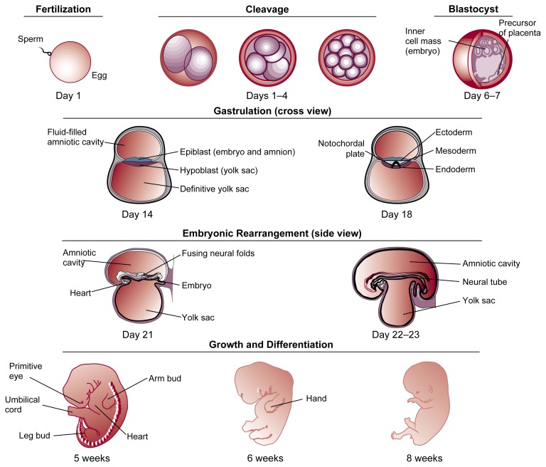
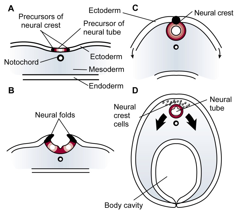
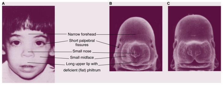
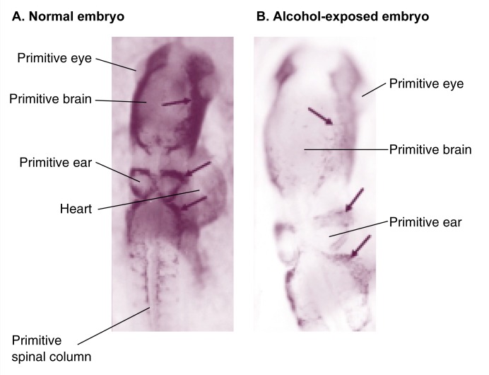
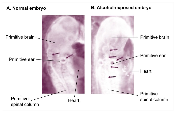
Exposure to alcohol during gestation can have profound consequences, but not all cells within the embryo are affected equally. Recent advances in molecular embryology have allowed an exploration of this variation. Much of this research has focused on the embryo's vulnerability to the facial malformations characteristic of fetal alcohol syndrome. Studies using mice and chicks show that alcohol exposure at specific stages of early embryo development results in significant death among the cells destined to give rise to facial structures (i.e., cranial neural crest cells). This type of cell death is through activation of the cell's own "self-destruct" machinery (i.e., apoptosis). Researchers have advanced several theories to explain how alcohol triggers apoptosis in the neural crest cells. These theories include deficiency in a type of vitamin A compound, retinoic acid; reduced levels of antioxidant compounds (i.e., free radical scavengers) that protect against damage from toxic oxygen molecules (i.e., free radicals); and interference with the cell's normal internal communication pathways.
Figures
Similar articles
-
Increased cell death and reduced neural crest cell numbers in ethanol-exposed embryos: partial basis for the fetal alcohol syndrome phenotype.Alcohol Clin Exp Res. 1995 Apr;19(2):378-86. doi: 10.1111/j.1530-0277.1995.tb01519.x. Alcohol Clin Exp Res. 1995. PMID: 7625573
-
The chick embryo as a model for the effects of prenatal exposure to alcohol on craniofacial development.Dev Biol. 2016 Jul 15;415(2):314-325. doi: 10.1016/j.ydbio.2016.01.007. Epub 2016 Jan 14. Dev Biol. 2016. PMID: 26777098 Review.
-
Neural crest development in fetal alcohol syndrome.Birth Defects Res C Embryo Today. 2014 Sep;102(3):210-20. doi: 10.1002/bdrc.21078. Epub 2014 Sep 15. Birth Defects Res C Embryo Today. 2014. PMID: 25219761 Free PMC article. Review.
-
Fetal alcohol syndrome and DiGeorge anomaly: critical ethanol exposure periods for craniofacial malformations as illustrated in an animal model.Am J Med Genet Suppl. 1986;2:97-112. doi: 10.1002/ajmg.1320250614. Am J Med Genet Suppl. 1986. PMID: 3146306
-
Genetic influences on craniofacial outcome in an avian model of prenatal alcohol exposure.Alcohol Clin Exp Res. 2001 Jan;25(1):60-9. Alcohol Clin Exp Res. 2001. PMID: 11198716
Cited by
-
Ethanol Attenuates Histiotrophic Nutrition Pathways and Alters the Intracellular Redox Environment and Thiol Proteome during Rat Organogenesis.Toxicol Sci. 2015 Oct;147(2):475-89. doi: 10.1093/toxsci/kfv145. Epub 2015 Jul 15. Toxicol Sci. 2015. PMID: 26185205 Free PMC article.
-
Transgenerational effects of binge drinking in a primate model: implications for human health.Fertil Steril. 2015 Feb;103(2):560-9. doi: 10.1016/j.fertnstert.2014.10.051. Epub 2014 Dec 6. Fertil Steril. 2015. PMID: 25492684 Free PMC article.
-
Effects of ethanol on physiological retinoic acid levels.IUBMB Life. 2011 Sep;63(9):701-6. doi: 10.1002/iub.500. Epub 2011 Jul 15. IUBMB Life. 2011. PMID: 21766417 Free PMC article. Review.
-
Ethanol, acetaldehyde, and estradiol affect growth and differentiation of rhesus monkey embryonic stem cells.Alcohol Clin Exp Res. 2011 Aug;35(8):1534-40. doi: 10.1111/j.1530-0277.2011.01490.x. Epub 2011 Mar 25. Alcohol Clin Exp Res. 2011. PMID: 21438889 Free PMC article.
-
Novel Ethanol-Sensitive Mutants Identified in an F3 Forward Genetic Screen.Alcohol Clin Exp Res. 2020 Jan;44(1):56-65. doi: 10.1111/acer.14240. Epub 2019 Dec 17. Alcohol Clin Exp Res. 2020. PMID: 31742718 Free PMC article.
References
Bibliography
-
- Gilbert SF. Developmental Biology. 2d ed. Sunderland, MA: Sinauer Associates; 1988.
-
- Gilbert SF, Raunio AM, editors. Embryology: Constructing the Organism. Sunderland, MA: Sinauer Associates; 1997.
-
- Larsen WJ. Essentials of Human Embryology. New York: Churchill Livingstone; 1998.
References
-
- Astley SJ, Clarren SK. A case definition and photographic screening tool for the facial phenotype of fetal alcohol syndrome. Journal of Pediatrics. 1996;129(1):33–41. - PubMed
-
- Bannigan J, Burke P. Ethanol teratogenicity in mice: A light microscopic study. Teratology. 1982;26(3):247–254. - PubMed
-
- Cartwright MM, Smith SM. Stage-dependent effects of ethanol on cranial neural crest cell development: Partial basis for the phenotypic variations observed in fetal alcohol syndrome. Alcoholism: Clinical and Experimental Research. 1995;19(6):1454–1462. - PubMed
-
- Cartwright MM, Tessmer LL, Smith SM. Ethanol-induced neural crest apoptosis is coincident with their endogenous death, but is mechanistically distinct. Alcoholism: Clinical and Experimental Research. 1998;22(1):142–149. - PubMed
-
- Chen S, Sulik KK. Free radicals and ethanol-induced cytotoxicity in neural crest cells. Alcoholism: Clinical and Experimental Research. 1996;20(6):1071–1076. - PubMed
Publication types
MeSH terms
Substances
LinkOut - more resources
Full Text Sources