. 2005 Jun 1;388(Pt 2):705-12.
doi: 10.1042/BJ20041966.

DNA-dependent phosphorylation of Chk1 and Claspin in a human cell-free system

Affiliations

DNA-dependent phosphorylation of Chk1 and Claspin in a human cell-free system

Catriona A L Clarke et al. Biochem J. .

Abstract

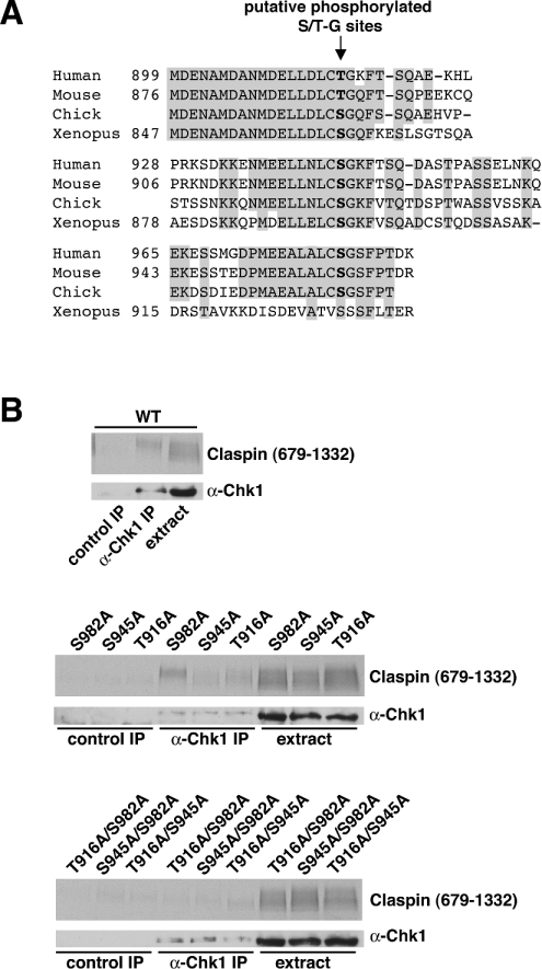
Cell-cycle checkpoints induced by DNA damage or replication play critical roles in the maintenance of genomic integrity during cell proliferation. Biochemical analysis of checkpoint pathways has been greatly facilitated by the use of cell-free systems made from Xenopus eggs. In the present study, we describe a human cell-free system that reproduces a DNA-dependent checkpoint pathway acting on the Chk1 protein kinase. In this system, double-stranded DNA oligonucleotides induce the phosphorylation of Chk1 at activating sites targeted by ATR [ATM (ataxia telangiectasia mutated)- and Rad3-related] and ATM kinases. Phosphorylation of Chk1 is dependent on the interaction of Claspin, a protein first identified in Xenopus as a Chk1-binding protein. We show that the DNA-dependent binding of Chk1 to Claspin requires two phosphorylation sites, Thr916 and Ser945, which lie within the Chk1-binding domain of Claspin. Using a phosphopeptide derived from the consensus motif of these sites, we show that the interaction of Claspin with Chk1 is required for the ATR/ATM-dependent phosphorylation of Chk1. Using a panel of protein kinase inhibitors, we provide evidence that Chk1 is phosphorylated at an additional site in response to activation of the checkpoint response, probably by autophosphorylation. Claspin is phosphorylated in the Chk1-binding domain in an ATR/ATM-dependent manner and is also targeted by additional kinases in response to double-stranded DNA oligonucleotides. This cell-free system will facilitate further biochemical analysis of the Chk1 pathway in humans.


Figures

Figure 1
Figure 1. Chk1 is phosphorylated at activating sites in a human cell-free system in response to double-stranded oligonucleotides
(A) Phosphorylation of Chk1 in HeLa cell extracts. HeLa cytoplasmic extract (S100), nuclear extract (HNE) and a mixture of equal volumes of the two (HNE/S100) were incubated at 30 °C for 30 min with or without an ATP-regenerating system, 10 μM OA and 50 ng/μl poly(dA/dT)70, as indicated. One sample contained 10 mM EDTA to block kinase activity. Western blots of protein separated by gel electrophoresis were probed with antibodies to Chk1 (anti-Chk1) or the phosphorylated Ser345 site in Chk1 (α-p345). (B) Time course of Chk1 phosphorylation. The extract was incubated with OA and poly(dA/dT)70 for the times shown and blotted with antibodies to Chk1 (anti-Chk1). (C) Chk1 is phosphorylated on Ser345 in response to dsDNA, not single-stranded DNA. The extract was incubated with OA and poly(dA/dT)70, poly(dA)70, poly(dT)70 or poly(dA/dT)40 as indicated, then Western blotted with either anti-Chk1 or α-p345. (D) Phosphorylation of Chk1 on Ser296 and Ser317 in response to oligonucleotides. The extract was incubated with OA and poly(dA/dT)70 as indicated and Western blotted with antibodies to Chk1 (anti-Chk1) and the phosphorylated Ser296 (α-p296) and Ser317 (α-p317) sites in Chk1.
Figure 2
Figure 2. Human Claspin is phosphorylated within its C-terminal half in response to double-stranded oligonucleotides
(A) Phosphorylation of endogenous Claspin. The extract was incubated with OA and poly(dA/dT)70 as indicated and blotted with antibodies to Chk1 (anti-Chk1), the phosphorylated Ser345 site in Chk1 (α-p345) and Claspin (anti-Claspin). (B) Phosphorylation of recombinant Claspin within residues 679–1332. The extract was incubated with radiolabelled fulllength Claspin, Claspin1–678 or Claspin679–1332, with or without OA and poly(dA/dT)70 as indicated. Proteins were detected by autoradiography. (C) Dephosphorylation of recombinant Claspin. The extract was incubated with radiolabelled His6-tagged full-length Claspin or Claspin679–1332, with or without OA+poly(dA/dT)70, as indicated. Proteins were precipitated, incubated further with or without protein phosphatase (λ-PPase), separated by SDS/PAGE and detected by autoradiography.
Figure 3
Figure 3. Phosphorylated Claspin co-precipitates with Chk1
The extract was incubated with radiolabelled full-length Claspin (1–1332), Claspin679–1332 or Claspin1–678, with or without OA and poly(dA/dT)70 as indicated, then precipitated (IP) with Protein G Dynal beads bound to either anti-Chk1 or control antibodies (both sheep). Beads were washed and proteins were boiled off the beads and analysed by autoradiography or blotting for Chk1 using a mouse monoclonal antibody. Extracts before precipitation were also analysed.
Figure 4
Figure 4. Thr916 and Ser945 in Claspin are essential for the binding interaction with Chk1
(A) Alignment of putative Chk1-binding domains in Claspin homologues from different vertebrates. Regions where the sequence is conserved between three species (including either serine or threonine residues) are shaded. The position of aligned putative Ser-Gly or Thr-Gly phosphorylation sites is indicated. (B) Mutational analysis of the putative Chk1-binding domain in human Claspin. Claspin679–1332 (WT, upper panel) and mutants in which each of the three putative phosphorylation sites, Thr916, Ser946 and Ser982, were changed to alanine (T916A, S945A and S982A) as single (middle panel) or double (lower panel) mutants were incubated with extract, OA and poly(dA/dT)70. Immunoprecipitations with anti-Chk1 or control sheep antibodies were performed and bound proteins were analysed by blotting for Chk1 with a mouse monoclonal antibody (anti-Chk1) or by autoradiography for Claspin679–1332.
Figure 5
Figure 5. A phosphopeptide derived from the Chk1-binding motif of Claspin disrupts binding and inhibits phosphorylation of Chk1
(A) Chk1-binding motif consensus peptide. Human (residues 908–923 and 937–952) and Xenopus (residues 856–871 and 887–102) repeat Chk1-binding motifs are aligned with a synthetic peptide consensus sequence. The serine residue replaced by an alanine or phosphorylated is indicated by an asterisk. (B) Disruption of the interaction between Claspin and Chk1. The extract was incubated with [35S]Claspin679–1332, OA and poly(dA/dT)70, with further additions of buffer or 1 μg/μl phosphoserine peptide (PS), alanine peptide (AS), serine peptide (SS) or an unrelated control peptide. Proteins precipitating with sheep anti-Chk1 or control antibodies were analysed by immunoblotting with mouse anti-Chk1 or autoradiography. (C) Inhibition of Chk1 phosphorylation. The extract was incubated with OA, poly(dA/dT)70 and different concentrations of PS (top panels), AS (middle panels) or SS (bottom panels). Samples were analysed by Western blotting with anti-Chk1 and α-p345 antibodies.
Figure 6
Figure 6. Effects of kinase inhibitors on Claspin and Chk1 phosphorylation
The extract was incubated with radiolabelled Claspin679–1332, radiolabelled GST-CKBD, OA+poly(dA/dT)70 and the following additions: staurosporine (staur), UCN01, roscovitine (rosco) and caffeine (upper panels); Gö6976, Ro318220, Bim1, SB203580, rapamycin (Rapa), PD98059 and H89 (lower panels). Proteins were detected by immunoblotting with antibodies raised against Chk1 (anti-Chk1) or specific phosphorylation sites on Chk1 (α-p345, α-p317 and α-p296). Claspin679–1332 and GST–CKBD proteins were detected by autoradiography.

References

    1. O'Connell M. J., Walworth N. C., Carr A. M. The G2-phase DNA-damage checkpoint. Trends Cell Biol. 2000;10:296–303. - PubMed
    1. Melo J., Toczyski D. A unified view of the DNA-damage checkpoint. Curr. Opin. Cell Biol. 2002;14:237–245. - PubMed
    1. Walworth N., Davey S., Beach D. Fission yeast chk1 protein kinase links the rad checkpoint pathway to cdc2. Nature (London) 1993;363:368–371. - PubMed
    1. Furnari B., Rhind N., Russell P. Cdc25 mitotic inducer targeted by chk1 DNA damage checkpoint kinase. Science. 1997;277:1495–1497. - PubMed
    1. Peng C. Y., Graves P. R., Thoma R. S., Wu Z., Shaw A. S., Piwnica-Worms H. Mitotic and G2 checkpoint control: regulation of 14-3-3 protein binding by phosphorylation of Cdc25C on serine-216. Science. 1997;277:1501–1505. - PubMed

Publication types