. 2005 Mar;79(5):2659-65.
doi: 10.1128/JVI.79.5.2659-2665.2005.

Roles of tumor necrosis factor alpha (TNF-alpha) and the p55 TNF receptor in CD1d induction and coxsackievirus B3-induced myocarditis

Affiliations

Roles of tumor necrosis factor alpha (TNF-alpha) and the p55 TNF receptor in CD1d induction and coxsackievirus B3-induced myocarditis

S A Huber et al. J Virol. 2005 Mar.

Abstract

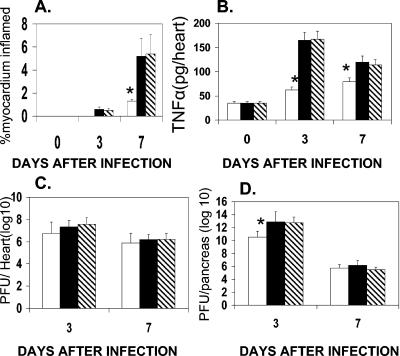
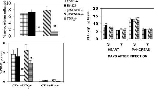
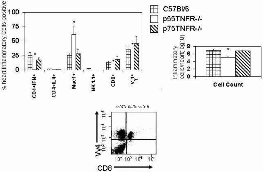
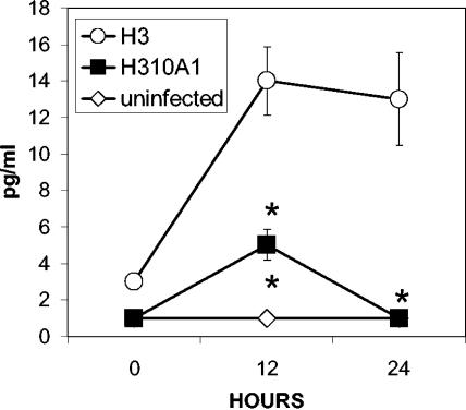
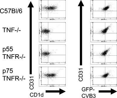
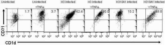
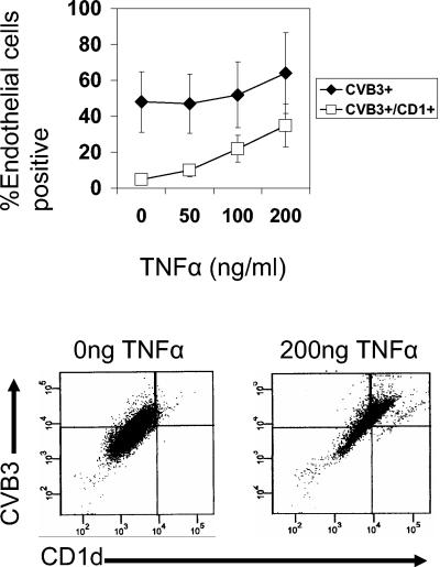
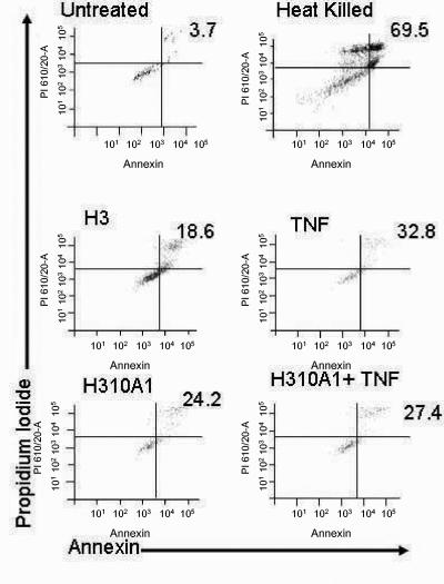
Giving C57BL/6 mice 10(4) PFU of coxsackievirus B3 (H3 variant) fails to induce myocarditis, but increasing the initial virus inoculum to 10(5) or 10(6) PFU causes significant cardiac disease. Virus titers in the heart were equivalent at days 3 and 7 in mice given all three virus doses, but day 3 titers in the pancreases of mice inoculated with 10(4) PFU were reduced. Tumor necrosis factor alpha (TNF-alpha) concentrations in the heart were increased in all infected mice, but cytokine levels were highest in mice given the larger virus inocula. TNF-alpha(-/-) and p55 TNF receptor-negative (TNFR(-/-)) mice developed minimal myocarditis compared to B6;129 or C57BL/6 control mice. p75 TNFR(-/-) mice were as disease susceptible as C57BL/6 animals. No significant differences in virus titers in heart or pancreas were observed between the groups, but C57BL/6 and p75 TNFR(-/-) animals showed 10-fold more inflammatory cells in the heart than p55 TNFR(-/-) mice, and the cell population was comprised of high concentrations of CD4(+) gamma interferon-positive and Vgamma4(+) cells. Cardiac endothelial cells isolated from C57BL/6 and p75 TNFR(-/-) mice upregulate CD1d, the molecule recognized by Vgamma4(+) cells, but infection of TNF(-/-) or p55 TNFR(-/-) endothelial cells failed to upregulate CD1d. Infection of C57BL/6 endothelial cells with a nonmyocarditic coxsackievirus B3 variant, H310A1, which is a poor inducer of TNF-alpha, failed to elicit CD1d expression, but TNF-alpha treatment of H310A1-infected endothelial cells increased CD1d levels to those seen in H3-infected cells. TNF-alpha treatment of uninfected endothelial cells had only a modest effect on CD1d expression, suggesting that optimal CD1d upregulation requires both infection and TNF-alpha signaling.


Figures

FIG. 1.
FIG. 1.
Myocarditis (A), TNF-α concentrations (B), cardiac virus titers (C), and pancreas virus titers (D) in C57BL/6 mice infected with 104 (open bars), 105 (black bars) or 106 (hatched bars) PFU of virus. Results are means ± standard errors of the means for four mice per group in one of three replicate experiments. *, significantly different from result for 106 PFU (P ≤ 0.05).
FIG. 2.
FIG. 2.
C57BL/6 and transgenic mice were infected with 105 PFU of H3 virus and killed 3 or 7 days later. Hearts were evaluated for myocarditis and virus titers. Peripheral blood mononuclear cells were labeled for CD4 and intracellular IFN-γ and IL-4 as described in Materials and Methods. Results are given as means ± standard errors of the means. *, significantly different from result for C57BL/6 (p55 TNFR−/−) or B6;129 (TNF-α−/−) mice (P < 0.05).
FIG. 3.
FIG. 3.
Infiltrating cells in the hearts of H3-infected C57BL/6, p55 TNFR−/−, and p75 TNFR−/− mice. Animals were infected with 105 PFU of virus and killed 7 days later. Hearts were perfused with PBS, then infiltrating cells were isolated and evaluated for the markers indicated. The total inflammatory cells isolated per heart for the three strains are shown. Results represent means ± standard errors of the means for four mice per group. *, significantly different from result for C57BL/6 mice (P < 0.05).
FIG. 4.
FIG. 4.
Hematoxylin-and-eosin-stained sections of myocardium from mice infected with 105 PFU of H3 virus. (A) C57BL/6 mice; (B) B6;129 mice; (C) TNF−/− mice; (D) p55 TNFR−/− mice; (E) p75 TNFR−/− mice.
FIG. 5.
FIG. 5.
TNF-α induction in endothelial cells infected with virus at an MOI of 100 and cultured for up to 24 h. Results are means ± standard errors of the means. *, significantly different from result for H3-infected (P ≤ 0.05).
FIG. 6.
FIG. 6.
Endothelial cells were cultured from the hearts of 6-week-old C57BL/6 and transgenic mice as indicated. Cells were infected with GFP-H3 virus at an MOI of 100 for 12 h and then labeled with PE-anti-CD1d and Cy-chrome-anti-CD31.
FIG. 7.
FIG. 7.
C57BL/6 cardiac endothelial cells were infected with either H3 or H310A1 virus at an MOI of 100 or were uninfected. Half of the cultures were treated with 200 ng of recombinant TNF-α per ml. Cells were labeled with Cy-chrome-anti-CD31 and PE-anti-CD1d at 12 h. Numbers in the upper right corners are the percentages of double-positive (CD31+ CD1d+) cells.
FIG. 8.
FIG. 8.
C57BL/6 cardiac endothelial cells were infected with H310A1 virus at an MOI of 100 and treated with between 0 and 200 ng of TNF-α per ml for 24 h. Cells were labeled with antibody to CD1d and CD31 (endothelial cell marker), fixed with 2% paraformaldehyde, and labeled with antibody to CVB3. The upper graph indicates the percentage of CD31+ cells which are also positive for CD1d and CVB3. Error bars indicate standard errors of the means. The lower graphs were gated on the CD31+ population and demonstrate that CD1d+ cells are also CVB3+ when treated with 200 ng of TNF-α per ml.
FIG. 9.
FIG. 9.
Dead cells in virus-infected and TNF-α-treated endothelial cell cultures. Endothelial cells were infected and/or treated with 200 ng of TNF-α per ml for 24 h. Cells were labeled with annexin V and propidium iodide to demonstrate dead cells (annexin V+ PI+). The numbers in the upper right quadrants indicate the percentages of dead cells.

Similar articles

Cited by

References

    1. Bergelson, J. M., J. G. Mohanty, R. L. Crowell, N. F. St. John, D. M. Lublin, and R. W. Finberg. 1995. Coxsackievirus B3 adapted to growth in RD cells binds to decay-accelerating factor (CD55). J. Virol. 69:1903-1906. - PMC - PubMed
    1. Chen, S., M. Sega, and A. Agarwal. 2004. “Lumen digestion” technique for isolation of aortic endothelial cells from heme oxygenase-1 knockout mice. BioTechniques 37:84-86, 88-89. - PubMed
    1. Choi, J. J., C. F. Reich III, and D. S. Pisetsky. 2004. Release of DNA from dead and dying lymphocyte and monocyte cell lines in vitro. Scand J Immunol 60:159-166. - PubMed
    1. Davis, L. S., S. S. Patel, J. P. Atkinson, and P. E. Lipsky. 1988. Decay-accelerating factor functions as a signal transducing molecule for human T cells. J. Immunol. 141:2246-2252. - PubMed
    1. Exley, M., N. Bigley, O. Cheng, S. Tahir, S. Smiley, Q. Carter, H. Stills, M. Grusby, Y. Koezuka, M. Taniguchi, and S. Balk. 2001. CD1d-reactive T-cell activation leads to amelioration of disease caused by diabetogenic encephalomyocarditis virus. J. Leukoc. Biol. 69:713-718. - PubMed

Publication types

MeSH terms

LinkOut - more resources