. 2005 Mar;79(5):2768-79.
doi: 10.1128/JVI.79.5.2768-2779.2005.

Human cytomegalovirus protein US11 provokes an unfolded protein response that may facilitate the degradation of class I major histocompatibility complex products

Affiliations

Human cytomegalovirus protein US11 provokes an unfolded protein response that may facilitate the degradation of class I major histocompatibility complex products

Boaz Tirosh et al. J Virol. 2005 Mar.

Abstract

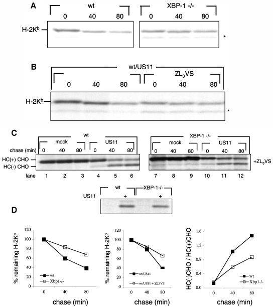
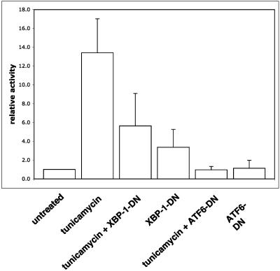
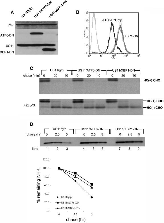
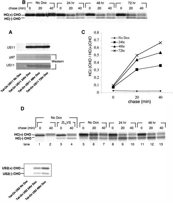
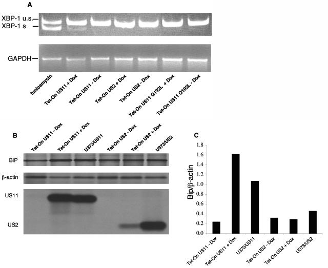
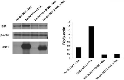
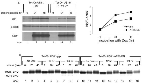
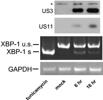
The human cytomegalovirus (HCMV) glycoprotein US11 diverts class I major histocompatibility complex (MHC) heavy chains (HC) from the endoplasmic reticulum (ER) to the cytosol, where HC are subjected to proteasome-mediated degradation. In mouse embryonic fibroblasts that are deficient for X-box binding protein 1 (XBP-1), a key transcription factor in the unfolded protein response (UPR) pathway, we show that degradation of endogenous mouse HC is impaired. Moreover, the rate of US11-mediated degradation of ectopically expressed HLA-A2 is reduced when XBP-1 is absent. In the human astrocytoma cell line U373, turning on expression of US11, but not US2, is sufficient to induce a UPR, as manifested by upregulation of the ER chaperone Bip and by splicing of XBP-1 mRNA. In the presence of dominant-negative versions of XBP-1 and activating transcription factor 6, the kinetics of class I MHC HC degradation were delayed when expression of US11 was turned on. The magnitude of these effects, while reproducible, was modest. Conversely, in cells that stably express high levels of US11, the degradation of HC is not affected by the presence of the dominant negative effectors of the UPR. An infection of human foreskin fibroblasts with human cytomegalovirus induced XBP-1 splicing in a manner that coincides with US11 expression. We conclude that the contribution of the UPR is more pronounced on HC degradation shortly after induction of US11 expression and that US11 is sufficient to induce such a response.


Figures

FIG. 1.
FIG. 1.
Class I MHC heavy chains are more stable in XBP-1−/− cells. (A) wt and XBP-1−/− MEF cells were pulse-labeled with [35S]methionine for 20 min and chased at 37°C up to 80 min. Cells were lysed in 1% SDS, and the lysate was then diluted to 0.07% SDS with NP-40 lysis mix followed by immunoprecipitation with anti-mouse H-2K serum (P8 antibody). The immunoprecipitates were analyzed by SDS-PAGE (12%). The background band is marked with an asterisk. (B) wt MEFs were transfected with US11 encoding pcDNA3, pulse-labeled with [35S]methionine for 20 min, and chased at 37°C up to 80 min in the presence or absence of the proteasome inhibitor ZL3VS. Cells were lysed in 1% SDS, and the lysate was then diluted to 0.07% SDS with NP-40 lysis mix followed by immunoprecipitation with anti-mouse H-2K serum (P8 antibody). The immunoprecipitates were analyzed by SDS-PAGE (12%). The background band is marked with an asterisk. (C) MEFs were transfected as described in Materials and Methods with an HLA-A2 encoding pcDNA3 with or without US11 encoding pcDNA3. Cells were pulse-labeled with [35S]methionine for 20 min and chased up to 80 min at 37°C in the presence of the proteasome inhibitor ZL3VS. Cells were lysed in 1% SDS, and the lysate was then diluted to 0.07% SDS with NP-40 lysis mix followed by immunoprecipitation with anti-class I heavy chain serum (αHC). US11 was sequentially immunoprecipitated from the zero time chase point (lower panel). Immunoprecipitates were analyzed by SDS-PAGE (12%). (D) Gels were quantified by phosphoimager.
FIG. 2.
FIG. 2.
Expression of XBP-1 and ATF6 dominant negatives attenuates tunicamycin-induced UPR. U373 cells were transfected as described in Materials and Methods with plasmids encoding firefly luciferase under the UPRE promoter and Renilla luciferase under thymidine kinase promoter. Plasmids encoding XBP-1-DN or ATF6-DN were cotransfected as indicated. Twenty-four hours after transfection, 1 μg of tunicamycin/ml was added for overnight treatment. Cells were harvested the next day and the dual-luciferase assay was performed. The results are averages from four independent experiments ± standard deviations.
FIG. 3.
FIG. 3.
US11-mediated degradation of HC is not affected by modulation of UPR in U373 cells. (A) Total cell lysates of U373 cells stably expressing XBP-1-DN-IRES-GFP, ATF6-DN-IRES-GFP, or an empty IRES-GFP construct were resolved by SDS-PAGE, and protein expression was examined by Western blotting. p97 was used as a loading control. (B) GFP levels were measured by flow cytometry. Untransfected U373 cells were measured as background. (C) U373 cell lines were pulse-labeled with [35S]methionine for 10 min and chased up to 40 min in the absence (upper panel) or presence (lower panel) of the proteasome inhibitor ZL3VS. Cells were lysed in 1% SDS, and the lysate was then diluted to 0.07% SDS with NP-40 lysis mix, followed by immunoprecipitation with αHC and analyzed by SDS-PAGE (12%). (D) U373 cell lines were transfected with pcDNA3 encoding the α1-AT NHK. Forty-eight hours after transfection, cells were pulse-labeled with [35S]methionine for 20 min and chased up to 5 h. Cells were lysed in NP-40 lysis buffer followed by immunoprecipitation with anti- α1-AT, and the immunoprecipitates were analyzed by SDS-PAGE (12%). Autoradiograms were quantified by phosphoimager.
FIG. 4.
FIG. 4.
MHC class I HC dislocation rate increases after US11 reaches steady state. (A) Tet-On US11 cells were incubated in the presence of DOX (1 μg/ml) for the indicated times. Cells were then harvested and divided into two portions. One-half of the samples were lysed, and US11 protein expression was examined by Western blotting. p97 was used as a loading control. (B) The second half of the samples were pulse-labeled with [35S]methionine for 10 min and chased for 40 min in the presence of the proteasome inhibitor ZL3VS. Cells were lysed in 1% SDS, and the lysate was then diluted to 0.07% SDS with NP-40 lysis mix followed by immunoprecipitation with αHC and analysis by SDS-12% PAGE. US11 was sequentially immunoprecipitated from the zero time point and similarly analyzed (A, upper panel). (C) Autoradiograms were quantified as mentioned above, and the deglycosylated HC to glycosylated HC ratio was then calculated. (D) Left, Tet-On US2 cells were incubated in the absence of DOX. Cells were pulse-labeled with [35S]methionine for 10 min and chased for 40 min in the presence or absence of the proteasome inhibitor ZL3VS. HC was immunoprecipitated as described for panel B. Immunoprecipitates were analyzed by SDS-PAGE (12%). Right, Tet-On US2 cells were incubated in the presence of DOX (1 μg/ml) for the indicated times. Cells were pulse-labeled with [35S]methionine for 10 min and chased for 40 min in the presence of the proteasome inhibitor ZL3VS. HC was immunoprecipitated as described for panel B. Lower panel, US2 was immunoprecipitated sequentially from the zero time chase point (right panel, lanes 5, 8, and 11). Immunoprecipitates were analyzed by SDS-PAGE (12%).
FIG. 5.
FIG. 5.
Expression of US11 triggers the UPR. (A) Tet-On cells were incubated for 48 h in the presence or absence of DOX (1 μg/ml). Total RNA was extracted, and XBP-1 splicing was analyzed by RT-PCR. PCR products were analyzed by 11% PAGE and visualized by ethidium bromide staining. (B) Tet-On cells and U373 cells that express US2 or US11 constitutively were pulse-labeled with [35S]methionine for 20 min. Cells were lysed in 1% SDS, and the lysate was then diluted to 0.07% SDS with NP-40 lysis mix. Samples were divided into three parts. One portion was immunoprecipitated with αKDEL monoclonal antibodies, the second portion was immunoprecipitated with anti-β-actin, and the third portion was immunoprecipitated with anti-US11 or anti-US2 antibodies. Loading was normalized to equal the number of trichloroacetic acid (TCA)-precipitable counts. (C) Autoradiograms were quantitated as mentioned above, and the ratio of Bip to β-actin was calculated.
FIG. 6.
FIG. 6.
The membrane-spanning domain of US11 mediates the induction of UPR. Tet-On cells for US11 or US11 Q192L mutant were incubated for 48 h in the presence or absence of DOX (1 μg/ml). Cells were pulse-labeled with [35S]methionine for 20 min. Cells were lysed in 1% SDS, and the lysate was then diluted to 0.07% SDS with NP-40 lysis buffer. Samples were divided into three parts. One portion was immunoprecipitated with αKDEL monoclonal antibodies, the second portion was immunoprecipitated with anti-β-actin, and the third portion was immunoprecipitated with anti-US11. Loading was normalized to equal the number of TCA-precipitable counts. (C) Autoradiograms were quantitated as mentioned above, and the ratio of Bip to β-actin was calculated.
FIG. 7.
FIG. 7.
Interference with the UPR attenuates the induction of Bip and decreases the dislocation of HC soon after turning on US11 expression. (A) Tet-On cells for US11 expressing either ATF6-DN or empty vector as the control were incubated for the indicated times in the presence or absence of DOX (1 μg/ml). Cells were pulse-labeled with [35S]methionine for 20 min. Cells were lysed in 1% SDS, and the lysate was then diluted to 0.07% SDS with NP-40 lysis buffer. Samples were divided into three parts. One portion was immunoprecipitated with αKDEL monoclonal antibodies, the second portion was immunoprecipitated with anti-β-actin, and the third portion was immunoprecipitated with anti-US11. Loading was normalized to equal the number of TCA-precipitable counts. Autoradiograms were quantitated as mentioned above, and the ratio of Bip to β-actin was calculated. (B) Cells were incubated for the indicated times with DOX. Cells were then harvested and pulse-labeled with [35S]methionine for 10 min and chased for 40 min in the presence of the proteasome inhibitor ZL3VS. Cells were lysed in 1% SDS, and the lysate was then diluted to 0.07% SDS with NP-40 lysis mix, followed by immunoprecipitation with αHC and analyzed by SDS-PAGE (12%).
FIG. 8.
FIG. 8.
UPR induction coincides with US11 expression upon HCMV infection. HFF cells were grown to 80% confluency and infected with HCMV at an MOI of 10. At the indicated times, cells were harvested. Ninety percent of the cells were used for RNA extraction and RT-PCR analysis of XBP-1 splicing as described for Fig. 5. The remainder of the samples were lysed in 1% SDS. Forty micrograms of total cell lysate was analyzed by Western blotting for the presence of US11 and US3.

Similar articles

Cited by

References

    1. Bonifacino, J. S., and A. M. Weissman. 1998. Ubiquitin and the control of protein fate in the secretory and endocytic pathways. Annu. Rev. Cell Dev. Biol. 14:19-57. - PMC - PubMed
    1. Casagrande, R., P. Stern, M. Diehn, C. Shamu, M. Osario, M. Zuniga, P. O. Brown, and H. Ploegh. 2000. Degradation of proteins from the ER of S. cerevisiae requires an intact unfolded protein response pathway. Mol. Cell. 5:729-735. - PubMed
    1. Ellgaard, L., and A. Helenius. 2001. ER quality control: towards an understanding at the molecular level. Curr. Opin. Cell Biol. 13:431-437. - PubMed
    1. Friedlander, R., E. Jarosch, J. Urban, C. Volkwein, and T. Sommer. 2000. A regulatory link between ER-associated protein degradation and the unfolded-protein response. Nat. Cell Biol. 2:379-384. - PubMed
    1. Furman, M. H., J. Loureiro, H. L. Ploegh, and D. Tortorella. 2003. Ubiquitinylation of the cytosolic domain of a type I membrane protein is not required to initiate its dislocation from the endoplasmic reticulum. J. Biol. Chem. 278:34804-34811. - PubMed

Publication types

MeSH terms