

Functional interaction of the adenovirus IVa2 protein with adenovirus type 5 packaging sequences
- PMID: 15709002
- PMCID: PMC548476
- DOI: 10.1128/JVI.79.5.2831-2838.2005
Functional interaction of the adenovirus IVa2 protein with adenovirus type 5 packaging sequences
Abstract
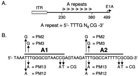
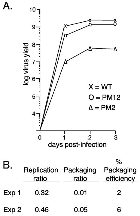
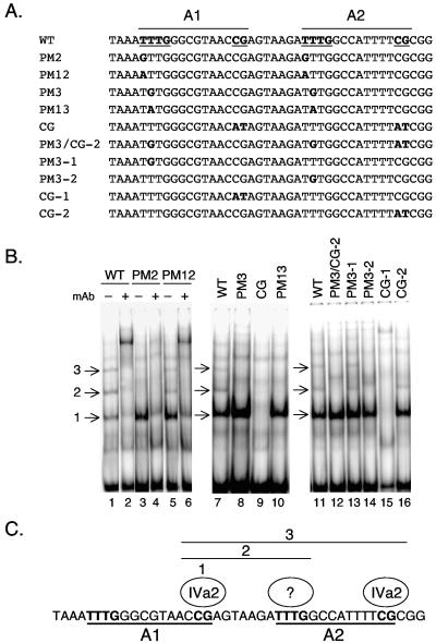
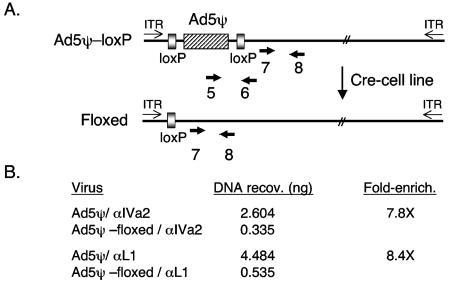
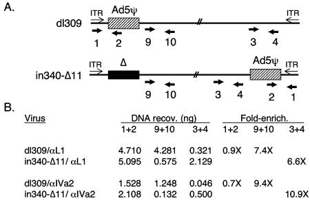
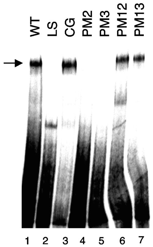
Adenovirus type 5 (Ad5) DNA packaging is initiated in a polar fashion from the left end of the genome. The packaging process is dependent on the cis-acting packaging domain located between nucleotides 230 and 380. Seven AT-rich repeats that direct packaging have been identified within this domain. A1, A2, A5, and A6 are the most important repeats functionally and share a bipartite sequence motif. Several lines of evidence suggest that there is a limiting trans-acting factor(s) that plays a role in packaging. Both cellular and viral proteins that interact with adenovirus packaging elements in vitro have been identified. In this study, we characterized a group of recombinant viruses that carry site-specific point mutations within a minimal packaging domain. The mutants were analyzed for growth properties in vivo and for the ability to bind cellular and viral proteins in vitro. Our results are consistent with a requirement of the viral IVa2 protein for DNA packaging via a direct interaction with packaging sequences. Our results also indicate that higher-order IVa2-containing complexes that form on adjacent packaging repeats in vitro are the complexes required for the packaging activity of these sites in vivo. Chromatin immunoprecipitation was used to study proteins that bind directly to the packaging sequences. These results demonstrate site-specific interaction of the viral IVa2 and L1 52/55K proteins with the Ad5 packaging domain in vivo. These results confirm and extend those previously reported and provide a framework on which to model the adenovirus assembly process.
Figures
Similar articles
-
Binding of CCAAT displacement protein CDP to adenovirus packaging sequences.J Virol. 2003 Jun;77(11):6255-64. doi: 10.1128/jvi.77.11.6255-6264.2003. J Virol. 2003. PMID: 12743282 Free PMC article.
-
Analysis of the interaction of the adenovirus L1 52/55-kilodalton and IVa2 proteins with the packaging sequence in vivo and in vitro.J Virol. 2005 Feb;79(4):2366-74. doi: 10.1128/JVI.79.4.2366-2374.2005. J Virol. 2005. PMID: 15681437 Free PMC article.
-
Cellular components interact with adenovirus type 5 minimal DNA packaging domains.J Virol. 1998 Aug;72(8):6339-47. doi: 10.1128/JVI.72.8.6339-6347.1998. J Virol. 1998. PMID: 9658073 Free PMC article.
-
Minimal cis-acting elements required for adenovirus genome packaging.J Virol. 2003 May;77(9):5127-35. doi: 10.1128/jvi.77.9.5127-5135.2003. J Virol. 2003. PMID: 12692215 Free PMC article.
-
Regulation of adenovirus packaging.Curr Top Microbiol Immunol. 2003;272:165-85. doi: 10.1007/978-3-662-05597-7_6. Curr Top Microbiol Immunol. 2003. PMID: 12747550 Review.
Cited by
-
Dependence of the encapsidation function of the adenovirus L1 52/55-kilodalton protein on its ability to bind the packaging sequence.J Virol. 2006 Feb;80(4):1965-71. doi: 10.1128/JVI.80.4.1965-1971.2006. J Virol. 2006. PMID: 16439552 Free PMC article.
-
The adenovirus L4-33K protein regulates both late gene expression patterns and viral DNA packaging.J Virol. 2013 Jun;87(12):6739-47. doi: 10.1128/JVI.00652-13. Epub 2013 Apr 3. J Virol. 2013. PMID: 23552425 Free PMC article.
-
Processing of the l1 52/55k protein by the adenovirus protease: a new substrate and new insights into virion maturation.J Virol. 2014 Feb;88(3):1513-24. doi: 10.1128/JVI.02884-13. Epub 2013 Nov 13. J Virol. 2014. PMID: 24227847 Free PMC article.
-
Localization of adenovirus morphogenesis players, together with visualization of assembly intermediates and failed products, favor a model where assembly and packaging occur concurrently at the periphery of the replication center.PLoS Pathog. 2017 Apr 27;13(4):e1006320. doi: 10.1371/journal.ppat.1006320. eCollection 2017 Apr. PLoS Pathog. 2017. PMID: 28448571 Free PMC article.
-
Cellular transcription factor TFII-I represses adenovirus gene expression.J Virol. 2025 Jun 17;99(6):e0061825. doi: 10.1128/jvi.00618-25. Epub 2025 May 12. J Virol. 2025. PMID: 40353670 Free PMC article.
References
Publication types
MeSH terms
Substances
Grants and funding
LinkOut - more resources
Full Text Sources
