. 2005 Mar;79(5):2931-40.
doi: 10.1128/JVI.79.5.2931-2940.2005.

The ectodomain of herpes simplex virus glycoprotein H contains a membrane alpha-helix with attributes of an internal fusion peptide, positionally conserved in the herpesviridae family

Affiliations

The ectodomain of herpes simplex virus glycoprotein H contains a membrane alpha-helix with attributes of an internal fusion peptide, positionally conserved in the herpesviridae family

Tatiana Gianni et al. J Virol. 2005 Mar.

Erratum in

  • J Virol. 2007 Mar;81(5):2539

Abstract

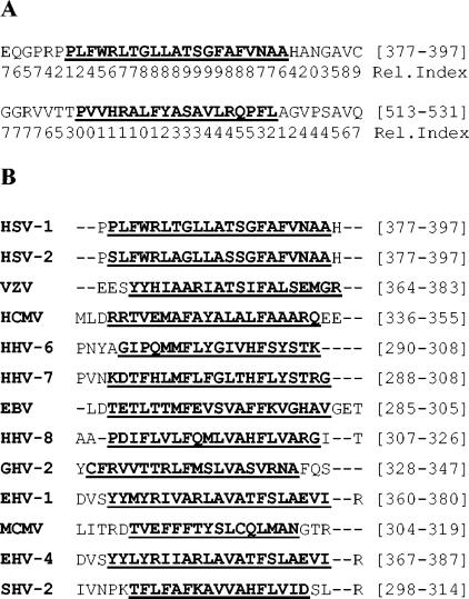
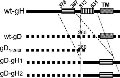
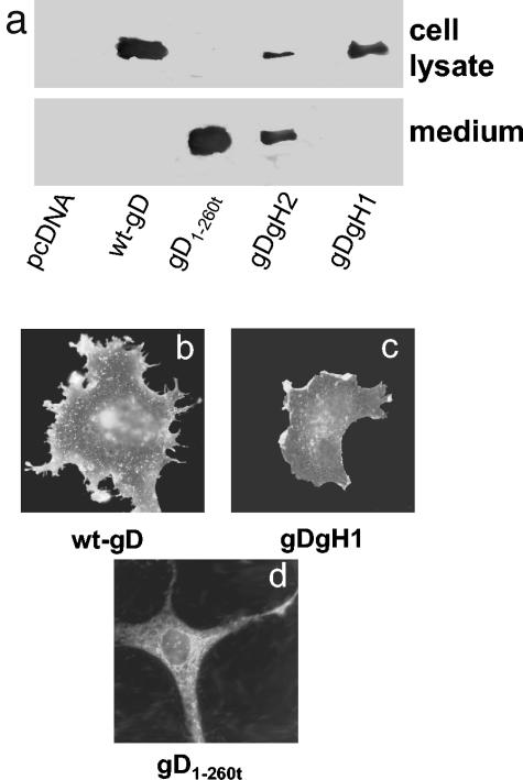
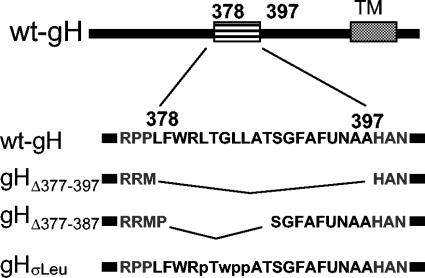
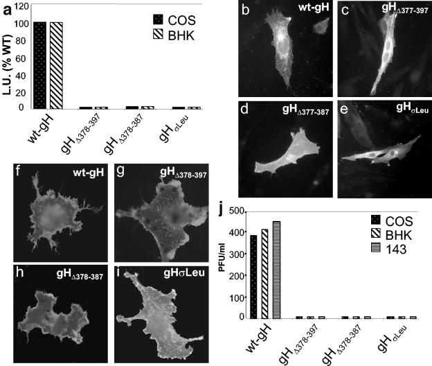
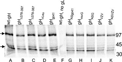
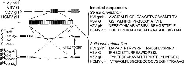
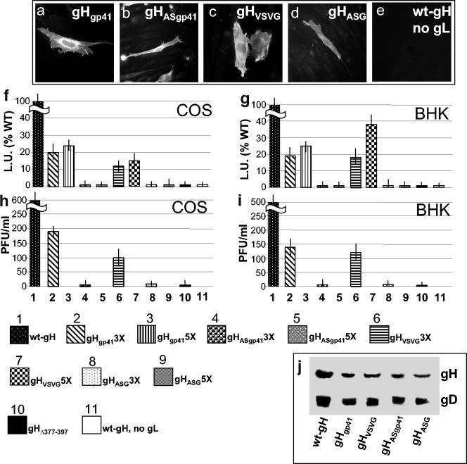
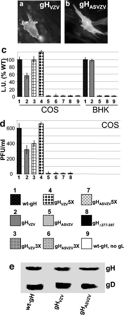
Human herpesviruses enter cells by fusion with target membranes, a process that requires three conserved glycoproteins: gB, gH, and gL. How these glycoproteins execute fusion is unknown. Neural network bioinformatics predicted a membrane alpha-helix contained within the ectodomain of herpes simplex virus (HSV) gH, positionally conserved in the gH of all examined herpesviruses. Evidence that it has attributes of an internal fusion peptide rests on the following lines of evidence. (i) The predicted membrane alpha-helix has the attribute of a membrane segment, since it transformed a soluble form of gD into a membrane-bound gD. (ii) It represents a critical domain of gH. Its partial or entire deletion, or substitution of critical residues inhibited HSV infectivity and fusion in the cell-cell fusion assay. (iii) Its replacement with the fusion peptide from human immunodeficiency virus gp41 or from vesicular stomatitis virus G partially rescued HSV infectivity and cell-cell fusion. The corresponding antisense sequences did not. (iv) The predicted alpha-helix located in the varicella-zoster virus gH ectodomain can functionally substitute the native HSV gH membrane alpha-helix, suggesting a conserved function in the human herpesviruses. We conclude that HSV gH exhibits features typical of viral fusion glycoproteins and that this property is likely conserved in the Herpesviridae family.


Figures

FIG. 1.
FIG. 1.
Prediction of membrane helices in gH sequences. (A) The two α-helices predicted in HSV-1 gH ectodomain by means of ENSEMBLE (24). Numbers in brackets indicate the first and last residue of the predicted helix. The reliability index (Rel.Index) evaluates the probability of correct prediction on a scale from 0 to 9. (B) Prediction of positionally conserved membrane α-helices in the gH of a number of herpesviruses. Numbers in brackets are as described for panel A. HHV, human herpesvirus; EBV, Epstein-Barr virus; GHV, gallid herpesvirus; EHV, equine herpesvirus; MCMV, murine cytomegalovirus; SHV, suid herpesvirus.
FIG. 2.
FIG. 2.
Schematic representation of the gD-gH1 and gD-gH2 constructs. The top line shows a wt-gH linear map with predicted α-helices and TM region. The second line shows a wt-gD linear map. An Asp718 site was inserted at residue 260 of gD; deletion of downstream sequences generated soluble gD1-260t (third line). Insertion of gH amino acid residues 354 to 416 and residues 505 to 551 at the C terminus of gD1-260t generated gD-gH1 and gD-gH2, respectively, is shown in the fourth and fifth lines.
FIG. 3.
FIG. 3.
Distribution of gD-gH1, gD-gH2, gD1-260t, and wt-gD and their cell surface expression. (a) Western blot analysis of the medium and cell lysates from COS cells transfected with plamids encoding gD1-260t, gD-gH1, gD-gH2, wt-gD, or pcDNA3.1. Cells and media were harvested 24 h after transfection. Western blot was developed with MAb H170 to gD. Note that the medium of cells expressing gD1-260t contains a secreted gD, whereas the medium of cells expressing gD-gH1 contains no gD and that of gD-gH2 contains some secreted gD; the lysates from cells expressing gD-gH1 accumulate higher quantities of gD than cells expressing gD-gH2 and gD1-260t. (b to d) IFA localization of gD in methanol-fixed COS cells expressing wt-gD (b), gD-gH1 (c), and gD1-260t (d) as detected with MAb H170. In panels b and c, the prominent localization of gD is at the cell surface. In panel d, the prominent localization of gD is reticular and diffuse to the cytoplasm.
FIG. 4.
FIG. 4.
Schematic representation of the gH deletion and mutation constructs. The top line shows a wt-gH linear map with predicted α-helix and TM regions. The second line shows the sequence of the α-helix, residues 378 to 397, and flanking residues. In gHΔ378-397 the α-helix was collapsed, after insertion of two SphI sites at residues 377 and 397. In gHΔ378-387 residues 378 to 387 were collapsed after insertion of SphI sites at residues 377 and 387. gHσLeu carries the L382P, GLL384-386WPP substitutions.
FIG. 5.
FIG. 5.
Cell fusion, cell surface expression, and infectivity complementation of gH mutants. (a) Quantification of luciferase-based cell fusion assay in effector cells transfected with plasmids encoding gL, gD, gB plus wt-gH, or the constructs gHΔ378-397, gHΔ378-387, and gHσLeu. The luciferase activity was expressed as luciferase units (L.U.). Each experiment was performed at least three times; the mean values are shown. (b to e) Paraformaldehyde-fixed permeabilized COS cells transfected with the indicated plasmid plus gL plasmid and stained with MAb 53S; reactivity denotes heterodimer formation with gL. (f to i) Paraformaldehyde-fixed COS cells transfected with the indicated plasmid plus gL, and stained with MAb 52S. (j) Infectivity complementation. The indicated cells were transfected with one of the gH plasmids and infected 4 h later with a gH−/+ stock of SCgHZ (7 PFU/cell). Progeny virus was titrated at 24 h in gH-expressing cells.
FIG. 6.
FIG. 6.
Coimmunoprecipitation of gH and gL by MAb 53S directed to gH. COS cells transfected with the indicated gH plasmid plus a gL plasmid were metabolically labeled with a mixture of [35S]methionine and [35S]cysteine for 16 h. gH was immunoprecipitated from the cell lysates with MAb 53S. gH coimmunoprecipitated gL in all samples. In lane F, in the absence of gL, MAb 53S failed to immunoprecipitate both gH and gL. On the right are indicated the relative migrations of molecular mass markers. Arrows on the left indicate the positions of gH and gL.
FIG. 7.
FIG. 7.
The top line shows a schematic linear map of HIV gp160, VSV-G, VZV gH, and HCMV gH; the HIV or VSV fusion peptides, or the predicted α-helix from VZV and HCMV gH, are marked as an enlarged box. Their sequences were cloned into the SphI site of gHΔ378-397, in sense or antisense directions. The right panel shows the sequence of the inserted fragments.
FIG. 8.
FIG. 8.
Expression, cell-cell fusion, and infectivity complementation of chimeric forms of gH. (a to e) BHK cells transfected with the indicated gH plasmid plus gL plasmid and reacted with MAb 53S. In panel e, gL plasmid was omitted. Positive reactivity indicates heterodimer formation with gL. (f and g) Quantification of luciferase-based cell-cell fusion assay in COS or BHK cells expressing the indicated gH plasmid plus gL, gD, and gB. The luciferase activity is expressed as luciferase units (L.U.). Figures represent percent values relative to wt-gH. Each experiment was performed at least three times. In lane 11, the transfection mixture contained wt-gH, gB, and gD plasmid and no gL plasmid. (h and i) Infectivity complementation, performed as described in legend to Fig. 5. Vertical bars represent the SD. (j) Quantification of gH and gD in complemented virions. Extracellular virions from the experiment in panel H were pelleted by high-speed centrifugation and analyzed by Western blotting with MAbs H12 to gH and H170 to gD. In the lane containing wt-gH a triple amount of virus was loaded.
FIG. 9.
FIG. 9.
Expression (a and b), cell-cell fusion (c), and infectivity complementation (d) of interspecific forms of gH, gHVZV, and gHAS-VZV. Details are as in the legend to Fig. 8. Vertical bars represent the SD. (e) Quantification of gH and gD in complemented virions, as detailed in Fig. 8J.

References

    1. Avitabile, E., G. Lombardi, and G. Campadelli-Fiume. 2003. Herpes simplex virus glycoprotein K, but not its syncytial allele, inhibits cell-cell fusion mediated by the four fusogenic glycoproteins, gD, gB, gH, and gL. J. Virol. 77:6836-6844. - PMC - PubMed
    1. Avitabile, E., G. Lombardi, T. Gianni, M. Capri, and G. Campadelli-Fiume. 2004. Coexpression of UL20p and gK inhibits cell-cell fusion mediated by herpes simplex virus glycoproteins gD, gH-gL, and wt-gB or an endocytosis-defective gB mutant, and downmodulates their cell surface expression. J. Virol. 78:8015-8025. - PMC - PubMed
    1. Bergeron, L., N. Sullivan, and J. Sodroski. 1992. Target cell-specific determinants of membrane fusion within the human immunodeficiency virus type 1 gp120 third variable region and gp41 amino terminus. J. Virol. 66:2389-2397. - PMC - PubMed
    1. Bressanelli, S., K. Stiasny, S. L. Allison, E. A. Stura, S. Duquerroy, J. Lescar, F. X. Heinz, and F. A. Rey. 2004. Structure of a flavivirus envelope glycoprotein in its low-pH-induced membrane fusion conformation. EMBO J. 23:728-738. - PMC - PubMed
    1. Bzik, D. J., B. A. Fox, N. A. DeLuca, and S. Person. 1984. Nucleotide sequence of a region of the herpes simplex virus type 1 gB glycoprotein gene: mutations affecting rate of virus entry and cell fusion. Virology 137:185-190. - PubMed

Publication types

MeSH terms

LinkOut - more resources