. 2005 Mar;79(5):3016-27.
doi: 10.1128/JVI.79.5.3016-3027.2005.

Immune tolerance split between hepatitis B virus precore and core proteins

Affiliations

Immune tolerance split between hepatitis B virus precore and core proteins

Margaret Chen et al. J Virol. 2005 Mar.

Abstract

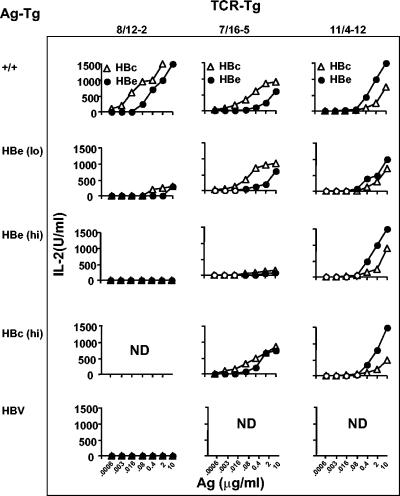
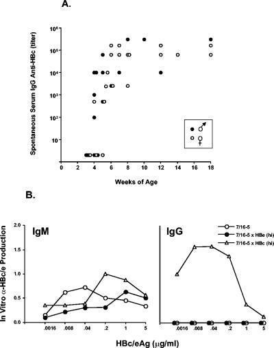
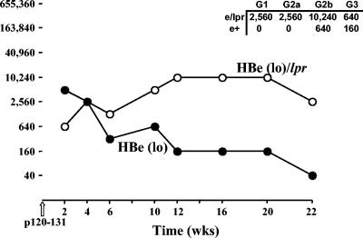
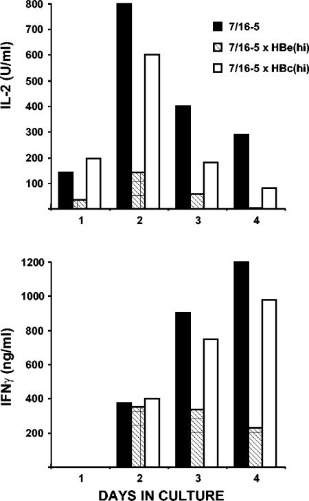
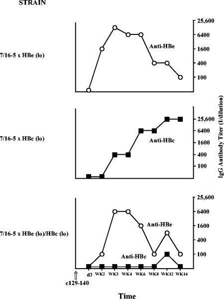
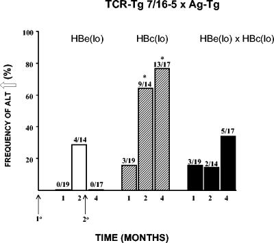
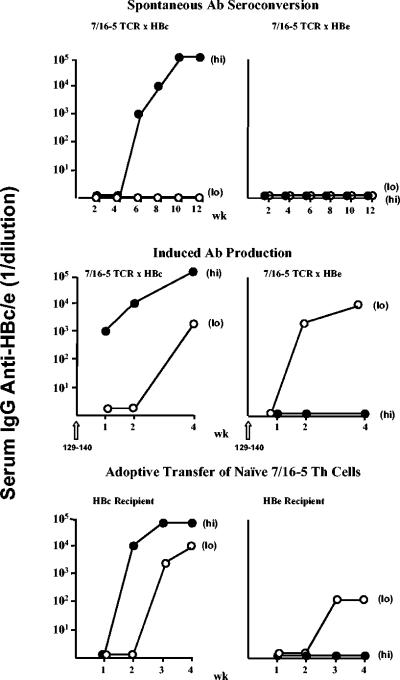
The function of the hepatitis B virus (HBV) precore or HBeAg is largely unknown because it is not required for viral assembly, infection, or replication. However, the HBeAg does appear to play a role in viral persistence. It has been suggested that the HBeAg may promote HBV chronicity by functioning as an immunoregulatory protein. As a model of chronic HBeAg exposure and to examine the tolerogenic potential of the HBV precore and core (HBcAg) proteins, HBc/HBeAg-transgenic (Tg) mice crossed with T cell receptor (TCR)-Tg mice expressing receptors for the HBc/HBeAgs (i.e., TCR-antigen double-Tg pairs) were produced. This study revealed three phenotypes of HBe/HBcAg-specific T-cell tolerance: (i) profound T-cell tolerance most likely mediated by clonal deletion, (ii) T-cell clonal ignorance, and (iii) nondeletional T-cell tolerance mediated by clonal anergy and dependent on the structure, location, and concentration of the tolerogen. The secreted HBeAg is significantly more efficient than the intracellular HBcAg at eliciting T-cell tolerance. The split T-cell tolerance between the HBeAg and the HBcAg and the clonal heterogeneity of HBc/HBeAg-specific T-cell tolerance may have significant implications for natural HBV infection and especially for precore-negative chronic hepatitis.


Figures

FIG. 1.
FIG. 1.
Summary of HBc/HBeAg-specific T-cell activation (IL-2 production) in TCR single-Tg and TCR × HBc or HBe double-Tg mice reveals the heterogeneity of HBeAg-specific T-cell tolerance. Unprimed, naive spleen cells from three TCR-Tg lineages (row 1) were cultured for 48 h with various concentrations of rHBcAg (HBc) and rHBeAg (HBe), and T-cell activation was determined by measuring IL-2 production. Rows 2 to 5 represent in vitro IL-2 production of naive T cells derived from TCR × Ag double-Tg pairs. The TCR-Tg (top) and the Ag-Tg (left side) lineages that comprise the double-Tg mice are depicted. The graphs represent the T-cell responses of single mice but are representative of the results from a minimum of six animals. ND, not determined; +/+, wild type.
FIG. 2.
FIG. 2.
Spontaneous anti-HBc seroconversion in 7/16-5 × HBc(hi) double-Tg mice (A) and comparative in vitro anti-HBc IgM and IgG antibody production in 7/16-5 TCR single-Tg and double-Tg mice (B). Individual double-Tg 7/16-5 × HBc(hi) female and male mice were bled beginning at 3 weeks of age and at intervals thereafter to determine the onset of anti-HBc IgG seroconversion and the persistence of anti-HBc antibodies (A). Serum IgG anti-HBc antibody titer (1/dilution) was determined by ELISA. Single HBc(hi)-Tg or single 7/16-5 TCR-Tg mice did not spontaneously produce anti-HBc antibody. (B) Unprimed naive spleen cells (5 × 10⁵/ml) from adult mice of the indicated 7/16-5 TCR single-Tg and 7/16-5 × HBe(hi) or 7/16-5 × HBc(hi) double-Tg strains were cultured with various concentrations of the rHBc/HBeAgs for 5 days. The 5-day supernatants were collected and assayed for IgM and IgG anti-HBc or anti-HBe antibodies by ELISA. The level of in vitro antibody production is expressed as an OD492 reading of undiluted supernatant in the antigen-specific ELISA. This experiment was performed on at least three separate occasions and is representative.
FIG. 3.
FIG. 3.
Comparative anti-HBe antibody production in B10.S HBe(lo) versus HBe(lo)/lpr single-Tg mice. Groups of five B10.S HBe(lo)-Tg or HBe(lo)/lpr-Tg mice were injected with the synthetic T-cell site p120-131 (50 μg) (incomplete Freund adjuvant). Mice were bleed at 2-week intervals, sera were pooled from five mice per group, and anti-HBe IgG antibody endpoint titers were determined by ELISA. Titers are expressed as the reciprocal of the highest dilution of sera to yield an OD492 value three times that of pre-p120-131 injection sera. wks, weeks.
FIG. 4.
FIG. 4.
Comparative kinetics of antigen-specific in vitro IL-2 and IFN-γ production by unprimed splenic T cells derived from 7/16-5 TCR single-Tg and 7/16-5 × HBe(hi) or 7/16-5 × HBc(hi) double-Tg mice. Unprimed spleen cells (5 × 10⁵/ml) from the indicated single- or double-Tg mice were cultured with the HBc/HBeAg-specific T-cell peptide p120-140 (0.5 μg/ml) for 4 days. Each day, culture supernatant was collected and analyzed for IL-2 or IFN-γ by ELISA. The data are representative of experiments performed a minimum of three times.
FIG. 5.
FIG. 5.
Comparative proliferative responses of 7/16-5 T cells derived from TCR single-Tg and 7/16-5 × HBe(hi) double-Tg mice. Unprimed spleen cells from 7/16-5 single-Tg or 7/16-5 × HBe(hi) double-Tg mice were labeled with the intracellular dye CFSE (0.5 mM) and cultured with 0.5 μg of the HBc/HBeAg-specific p120-140 for 4 or 7 days. Cultured cells were harvested at 4 or 7 days and labeled with anti-TCR Vβ11 antibodies. The number of cell divisions is measured by the dilution of the CFSE dye, which is reduced by 50% at each cell division, in TCR Vβ11+-gated T cells by flow cytometry. The numbers above the windows represent the percentage of Vβ11+ T cells that have undergone the indicated number of cell divisions. The data are representative of the results from two separate experiments.
FIG. 6.
FIG. 6.
Effect of serum HBeAg on the humoral and T-cell response to immunization with rHBcAg. (A) Groups of three adult B10.S wild-type (B10.S/+) and B10.S HBe(hi)-Tg mice were immunized with rHBcAg (10 μg) (incomplete Freund adjuvant), and IgG anti-HBc antibody was measured in pooled sera by ELISA at 2-week intervals. Anti-HBc antibody is expressed as an endpoint titer (1/dilution). (B) Spleen cells from the mice depicted in Fig. 6A were harvested 6 weeks after rHBcAg immunization and cultured (5 × 10⁶/ml) with various concentrations of rHBcAg for 4 days. Supernatants were collected and analyzed for IFN-γ. Comparative IFN-γ levels were expressed as an OD492 reading of the undiluted supernatants in the IFN-γ-specific ELISA. Data represent means ± standard deviations of the results for 3 mice per group. The experiment shown is representative of three performed. wk, week.
FIG. 7.
FIG. 7.
Perinatal exposure to the HBeAg is tolerogenic. Young (3 to 4 weeks of age [WOA]) B10.S wild-type (B10.S/+) or B10.S HBe(hi)-Tg mice, which were HBeAg negative at birth, were immunized with rHBeAg (10 μg) (incomplete Freund adjuvant). Ten days later, spleen cells were harvested and cultured with various concentrations of the dominant HBc/HBeAg-specific T-cell site in B10.S mice, p120-131. After 2 days of spleen culture, supernatants were collected and analyzed for IL-2 by ELISA. Comparative IL-2 levels were expressed as OD492 readings of undiluted supernatants in the IL-2-specific ELISA. The symbols represent individual mice. This experiment was performed on two occasions, and the results are representative.
FIG. 8.
FIG. 8.
Induction of low-level liver injury in 7/16-5 × HBc(lo) double-Tg mice. Two 7/16-5 × HBc(lo) double-Tg mice (A and B) were injected with the 7/16-5 TCR-specific peptide 129-140 (100 μg) (CFA), and at multiple intervals, sera were collected and analyzed for sALT and IgG anti-HBc antibodies. No liver injury occurred in 7/16-5 TCR single-Tg or HBcAg single-Tg mice injected with p129-140 (data not shown). d, day; WK, week.
FIG. 9.
FIG. 9.
Induced seroconversion in 7/16-5 TCR × HBc and HBeAg double-Tg and triple-Tg mice. Groups of three mice each of the indicated double- and triple-Tg strains were injected with the 7/16-5 TCR-specific peptide 129-140 (100 μg) (CFA), and sera were collected at multiple intervals, pooled, and analyzed for IgG anti-HBe and IgG anti-HBc antibodies by ELISA. Antibody titer is defined as the endpoint serum dilution to yield three times the OD492 value of equally diluted preimmunization sera. The results are representative of experiments performed on two separate occasions. d, day; WK, week.
FIG. 10.
FIG. 10.
Liver injury induced in 7/16-5 × HBc(lo) double-Tg mice is moderated by serum HBeAg. Groups of six 7/16-5 × HBe(lo) double-Tg, 7/16-5 × HBc(lo) double-Tg, and 7/16-5 × HBe(lo) × HBc(lo) triple-Tg mice were injected at time zero with p129-140 (100 μg) (CFA) and again at 2 months (50 μg) (incomplete Freund adjuvant). At multiple intervals, 2 to 3 mice from each group were bled and individual sera were analyzed for ALT elevation. sALT was considered elevated above 100 U/liter. Frequencies of sALT elevations in each group during months 1, 2, and 4 are shown. The numbers above the bars depict the absolute number of sera demonstrating heightened ALT values per total number of sera tested in a given time frame. *, P < 0.01 versus 7/16-5 × HBe(lo) and 7/16-5 × HBe(lo) × HBc(lo) groups (unpaired t test).
FIG. 11.
FIG. 11.
Summary of spontaneous anti-HBc/HBe antibody (Ab) seroconversion and induced antibody production in 7/16-5 × HBc and 7/16-5 × HBe double-Tg mice and antibody production after adoptive transfer of 7/16-5 T cells reveals the tolerance split between the HBeAg and the HBcAg. Spontaneous antibody seroconversion (top row) was monitored by bimonthly blood samples and measurement of IgG anti-HBc and anti-HBe antibodies by ELISA. “(Hi)” and “(Lo)” represent the antigen expression levels of HBc-Tg (left panels) and HBe-Tg (right panels) mice. Induced IgG antibody production in the indicated 7/16-5 double-Tg mice (middle row) was accomplished by injecting p129-140 (50 to 100 μg) (incomplete Freund adjuvant) at time zero and measuring anti-HBc or anti-HBe antibody production at 1, 2, and 4 weeks postinjection. In adoptive transfer experiments (bottom row), unprimed, naive spleen cells derived from 7/16-5 TCR-Tg mice were transferred (30 × 106 cells, intravenously) into CD4/CD8-depleted HBc-Tg or HBe-Tg recipients. Recipient sera were collected at weekly intervals and analyzed for anti-HBc and anti-HBe IgG antibodies by ELISA. The symbols in this figure represent individual mice; however, the results are representative of experiments performed on multiple occasions with a minimum of 6 mice per group. wk, week.

Similar articles

Cited by

References

    1. Arakawa, K., F. Tsuda, K. Takahashi, I. Ise, S. Naito, E. Kosugi, Y. Miyakawa, and M. Mayumi. 1982. Maternofetal transmission of IgG-bound hepatitis B e antigen. Pediatr. Res. 16:247-250. - PubMed
    1. Bertoletti, A., C. Ferrari, F. Fiaccadori, A. Penna, R. Margolskee, H. J. Schlicht, P. Fowler, and F. V. Chisari. 1991. HLA class I-restricted human cytotoxic T cells recognize endogenously synthesized hepatitis B virus nucleocapsid antigen. Proc. Natl. Acad. Sci. USA 88:10445-10449. - PMC - PubMed
    1. Brunetto, M. R., M. M. Giarin, F. Oliveri, E. Chiaberge, M. Baldi, A. Alfarano, A. Serra, G. Saracco, G. Verme, H. Will, and F. Bonino. 1991. Wild-type and e antigen-minus hepatitis B viruses and course of chronic hepatitis. Proc. Natl. Acad. Sci. USA 88:4186-4190. - PMC - PubMed
    1. Carman, W. F., M. R. Jacyna, S. Hadziyannis, P. Karayiannis, M. J. McGarvey, A. Makris, and H. C. Thomas. 1989. Mutation preventing formation of hepatitis B e antigen in patients with chronic hepatitis B infection. Lancet ii:588-590. - PubMed
    1. Carman, W. F., E. A. Fagan, S. Hadziyannis, P. Karayiannis, N. C. Tassopoulos, R. Williams, and H. C. Thomas. 1991. Association of a precore genomic variant of hepatitis B virus with fulminant hepatitis. Hepatology 14:219-222. - PubMed

Publication types