. 2005 Mar;79(5):3200-5.
doi: 10.1128/JVI.79.5.3200-3205.2005.

Entry of pseudorabies virus: an immunogold-labeling study

Affiliations

Entry of pseudorabies virus: an immunogold-labeling study

Harald Granzow et al. J Virol. 2005 Mar.

Abstract

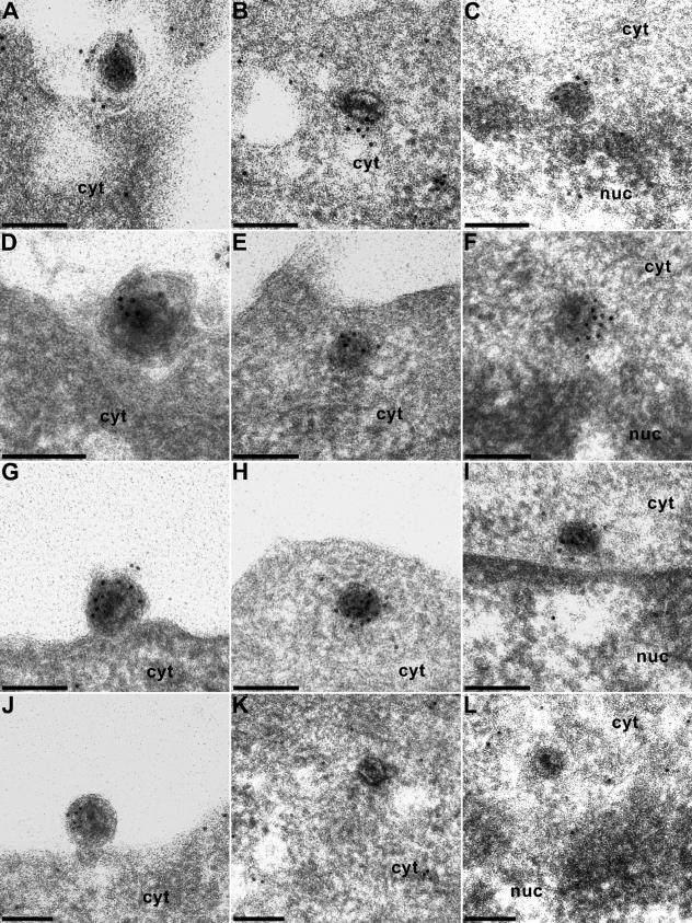
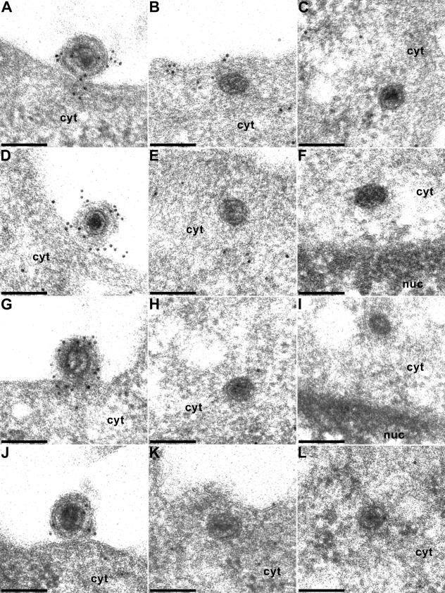
Herpesviruses infect cells by fusion of the viral envelope with cellular membranes, primarily the plasma membrane. During this process structural components of the mature virion are lost from the invading nucleocapsid, which then travels along microtubules to the nuclear pore. We examined the penetration process by immunoelectron microscopy and analyzed which of the major tegument proteins remained associated with the incoming capsid. We show that the UL36, UL37, and US3 proteins were present at intracytoplasmic capsids after penetration, whereas the UL11, UL47, UL48, and UL49 tegument proteins were lost. Thus, the three capsid-associated tegument proteins are prime candidates for viral proteins that interact with cellular motor proteins for transport of nucleocapsids to the nucleus.


Figures

FIG. 1.
FIG. 1.
Ultrastructure of the entry process of PrV. PrV virions were documented during attachment (A) and penetration (B), associated with microtubules in the cytoplasm (C and D), and at the nuclear pore (E and F). Bars, 150 (A and F), 200 (B to D), and 500 (E) nm.
FIG. 2.
FIG. 2.
Immunogold labeling of virions during entry. Extracellular PrV virions during attachment or fusion (A, D, G, and J), in the cytoplasm after penetration (B, E, H, and K), and at the nuclear pore (C, F, I, and L) were immunogold labeled with antisera to the US3 (A to C), UL36 (D to F), UL37 (G to I), and UL11 (J to L) proteins. cyt, cytoplasm; nuc, nucleus. Bars, 150 nm.
FIG. 3.
FIG. 3.
Immunogold labeling of virions during entry. Extracellular PrV virions during attachment or fusion (A, D, G, and J), in the cytoplasm after penetration (B, C, E, H, K, and L), and at the nuclear pore (F and I) were immunogold labeled with antisera to the UL46 (A to C), UL47 (D to F), UL48 (G to I), and UL49 (J to L) proteins. cyt, cytoplasm; nuc, nucleus. Bars, 150 nm.

References

    1. Baines, J. D., and B. Roizman. 1992. The UL11 gene of herpes simplex virus encodes a function that facilitates nucleocapsid envelopment and egress from cells. J. Virol. 66:5168-5174. - PMC - PubMed
    1. Batterson, W., and B. Roizman. 1983. Characterization of the herpes simplex virion-associated factor responsible for the induction of α-genes. J. Virol. 46:371-377. - PMC - PubMed
    1. Bowzard, J. B., R. J. Visalli, C. B. Wilson, J. S. Loomis, E. M. Callahan, R. J. Courtney, and J. W. Wills. 2000. Membrane targeting properties of a herpesvirus tegument protein-retrovirus Gag chimera. J. Virol. 74:8692-8699. - PMC - PubMed
    1. Diefenbach, R. J., M. Miranda-Saksena, E. Diefenbach, D. J. Holland, R. A. Boadle, P. J. Armati, and A. L. Cunningham. 2002. Herpes simplex virus tegument protein US11 interacts with conventional kinesin heavy chain. J. Virol. 76:3282-3291. - PMC - PubMed
    1. Döhner, K. A. Wolfstein, U. Prank, C. Echeverri, D. Dujardin, R. Vallee, and B. Sodeik. 2002. Function of dynein and dynactin in herpes simplex virus capsid transport. Mol. Biol. Cell 13:2795-2809. - PMC - PubMed

Publication types

LinkOut - more resources