

The coiled-coil domain of the adenovirus type 5 protein IX is dispensable for capsid incorporation and thermostability
- PMID: 15709043
- PMCID: PMC548437
- DOI: 10.1128/JVI.79.5.3206-3210.2005
The coiled-coil domain of the adenovirus type 5 protein IX is dispensable for capsid incorporation and thermostability
Abstract
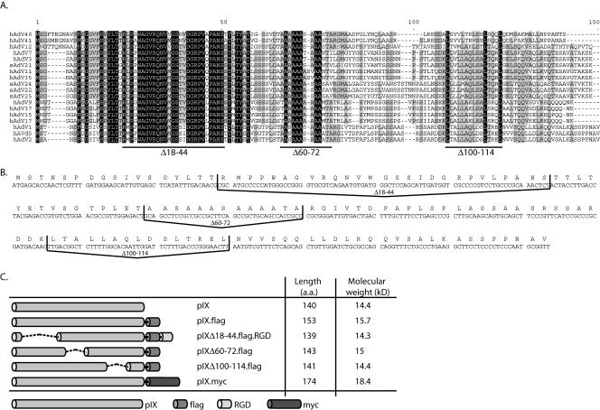
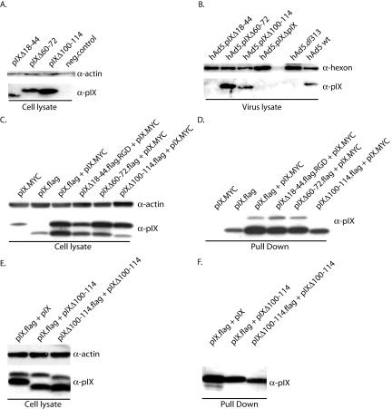
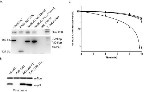
The 14.4-kDa hexon-associated protein IX (pIX) acts as a cement in the capsids of primate adenoviruses and confers a thermostable phenotype. Here we show that deletion of amino acids 100 to 114 of adenovirus type 5 pIX, which eliminates the conserved coiled-coil domain, impairs its capacity to self-associate. However, pIXDelta100-114 is efficiently incorporated into the viral capsid, and the resulting virions are thermostable. Deletion of the central alanine-rich domain, as in pIXDelta60-72, does not impair self-association, incorporation into the capsid, or the thermostable phenotype. These data demonstrate, first, that the self-association of pIX is dispensable for its incorporation into the capsid and generation of the thermostability phenotype and, second, that the increased thermostability results from pIX monomers binding to different hexon capsomers rather than capsid stabilization by pIX multimers.
Figures
Similar articles
-
Bovine adenovirus type 3 containing heterologous protein in the C-terminus of minor capsid protein IX.Virology. 2004 Mar 15;320(2):291-300. doi: 10.1016/j.virol.2003.12.007. Virology. 2004. PMID: 15016551
-
Comparison of the deduced amino acid sequence of guinea pig adenovirus hexon protein with that of other mastadenoviruses.Comp Med. 2001 Apr;51(2):120-6. Comp Med. 2001. PMID: 11922174
-
Genetic incorporation of HSV-1 thymidine kinase into the adenovirus protein IX for functional display on the virion.Virology. 2005 Aug 1;338(2):247-58. doi: 10.1016/j.virol.2005.04.005. Virology. 2005. PMID: 15996701
-
Adenovirus protein IX: a new look at an old protein.Mol Ther. 2005 Jan;11(1):19-25. doi: 10.1016/j.ymthe.2004.09.018. Mol Ther. 2005. PMID: 15585402 Review.
-
The adenovirus capsid: major progress in minor proteins.J Gen Virol. 2005 Jun;86(Pt 6):1581-1588. doi: 10.1099/vir.0.80877-0. J Gen Virol. 2005. PMID: 15914835 Review.
Cited by
-
Latest insights on adenovirus structure and assembly.Viruses. 2012 May;4(5):847-77. doi: 10.3390/v4050847. Epub 2012 May 21. Viruses. 2012. PMID: 22754652 Free PMC article. Review.
-
Cryoelectron microscopy of protein IX-modified adenoviruses suggests a new position for the C terminus of protein IX.J Virol. 2006 Dec;80(23):11881-6. doi: 10.1128/JVI.01471-06. Epub 2006 Sep 20. J Virol. 2006. PMID: 16987967 Free PMC article.
-
Near-atomic structure of an atadenovirus reveals a conserved capsid-binding motif and intergenera variations in cementing proteins.Sci Adv. 2021 Mar 31;7(14):eabe6008. doi: 10.1126/sciadv.abe6008. Print 2021 Mar. Sci Adv. 2021. PMID: 33789897 Free PMC article.
-
Transductional targeting of adenovirus vectors for gene therapy.Cancer Gene Ther. 2006 Sep;13(9):830-44. doi: 10.1038/sj.cgt.7700928. Epub 2006 Jan 27. Cancer Gene Ther. 2006. PMID: 16439993 Free PMC article. Review.
-
Cryo-EM structure of enteric adenovirus HAdV-F41 highlights structural variations among human adenoviruses.Sci Adv. 2021 Feb 24;7(9):eabd9421. doi: 10.1126/sciadv.abd9421. Print 2021 Feb. Sci Adv. 2021. PMID: 33627423 Free PMC article.
References
-
- Fallaux, F. J., O. Kranenburg, S. J. Cramer, A. Houweling, H. van Ormondt, R. C. Hoeben, and A. J. van der Eb. 1996. Characterization of 911: a new helper cell line for the titration and propagation of early region 1-deleted adenoviral vectors. Hum. Gene Ther. 7:215-222. - PubMed
Publication types
MeSH terms
Substances
LinkOut - more resources
Full Text Sources
Other Literature Sources
