

Diffusion-weighted imaging abnormalities after percutaneous transluminal angioplasty and stenting for intracranial atherosclerotic disease
- PMID: 15709141
- PMCID: PMC7974074
Diffusion-weighted imaging abnormalities after percutaneous transluminal angioplasty and stenting for intracranial atherosclerotic disease
Abstract
Background and purpose: The literature contains relatively few reports of distal embolism associated with intervention for intracranial atherosclerotic disease. Our purpose was to evaluate the frequency of thromboembolic events after percutaneous transluminal angioplasty (PTA) or stent placement in this setting by using diffusion-weighted (DW) imaging.
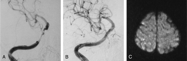
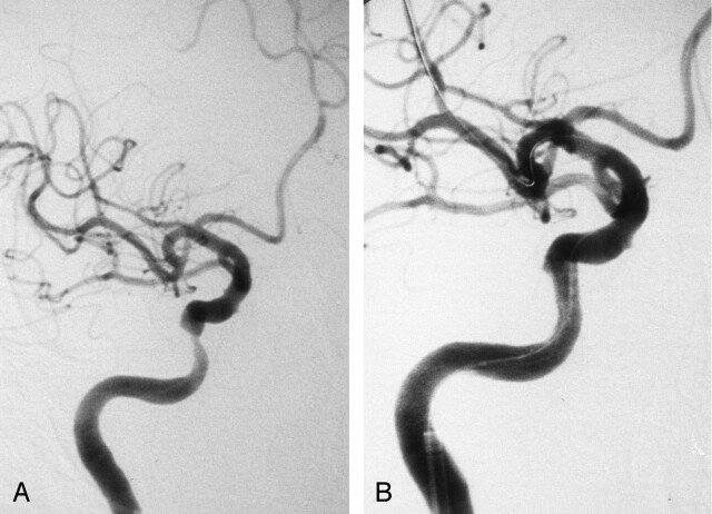
Methods: Between October 1999 and January 2004, 16 consecutive patients with symptomatic intracranial arterial stenosis greater than 60% were treated with PTA or stent placement without a protection system. Whole-brain DW imaging was performed before and after intervention. DW imaging findings were retrospectively analyzed and divided into three groups according to new hyperintensities: type A was none; type B, a single lesion; and type C, multiple lesions.
Results: Nine type A, five type B, and three type C lesions were detected after the interventions. All hyperintense lesions were less than 5 mm in diameter. All type C lesions occurred in the context of internal carotid artery stenosis treated with stent placement. DW imaging abnormalities occurred most frequently when PTA followed by stent placement was performed for long internal carotid artery stenoses. No new neurologic deficits occurred in any patient.
Conclusion: In this series, PTA or stent placement or both for intracranial atherosclerotic lesions was safe. New DW imaging abnormalities were less frequent in patients who underwent PTA alone or primary stent placement than in those receiving PTA followed by stent placement.
Figures
References
-
- Craig DR, Meguro K, Watridge C, et al. Intracranial internal carotid artery stenosis. Stroke 1982;13:825–828 - PubMed
-
- Arenillas JF, Molina CA, Montaner J, et al. Progression and clinical recurrence of symptomatic middle cerebral artery stenosis: a long-term follow-up transcranial Doppler ultrasound study. Stroke 2001;32:2898–2904 - PubMed
-
- Terada T, Tsuura M, Matsumoto H, et al. Endovascular therapy for stenosis of the petrous or cavernous portion of the internal carotid artery: percutaneous transluminal angioplasty compared with stent placement. J Neurosurg 2003;98:491–497 - PubMed
-
- Gomez CR, Misra VK, Liu MW, et al. Elective stenting of symptomatic basilar artery stenosis. Stroke 2000;31:95–99 - PubMed
MeSH terms
LinkOut - more resources
Full Text Sources