

Pseudomonas aeruginosa MutL protein functions in Escherichia coli
- PMID: 15709980
- PMCID: PMC1183468
- DOI: 10.1042/BJ20042073
Pseudomonas aeruginosa MutL protein functions in Escherichia coli
Abstract
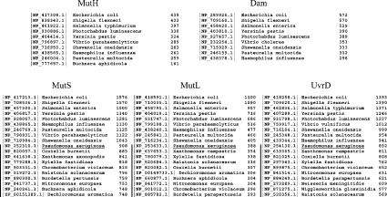
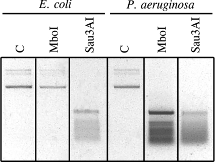
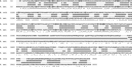
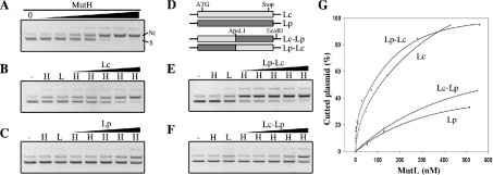
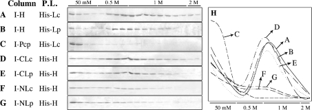
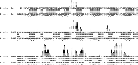
Escherichia coli MutS, MutL and MutH proteins act sequentially in the MMRS (mismatch repair system). MutH directs the repair system to the newly synthesized strand due to its transient lack of Dam (DNA-adenine methylase) methylation. Although Pseudomonas aeruginosa does not have the corresponding E. coli MutH and Dam homologues, and consequently the MMRS seems to work differently, we show that the mutL gene from P. aeruginosa is capable of complementing a MutL-deficient strain of E. coli. MutL from P. aeruginosa has conserved 21 out of the 22 amino acids known to affect functioning of E. coli MutL. We showed, using protein affinity chromatography, that the C-terminal regions of P. aeruginosa and E. coli MutL are capable of specifically interacting with E. coli MutH and retaining the E. coli MutH. Although, the amino acid sequences of the C-terminal regions of these two proteins are only 18% identical, they are 88% identical in the predicted secondary structure. Finally, by analysing (E. coli-P. aeruginosa) chimaeric MutL proteins, we show that the N-terminal regions of E. coli and P. aeruginosa MutL proteins function similarly, in vivo and in vitro. These new findings support the hypothesis that a large surface, rather than a single amino acid, constitutes the MutL surface for interaction with MutH, and that the N- and C-terminal regions of MutL are involved in such interactions.
Figures
Similar articles
-
In vitro and in vivo studies of MutS, MutL and MutH mutants: correlation of mismatch repair and DNA recombination.DNA Repair (Amst). 2003 Apr 2;2(4):387-405. doi: 10.1016/s1568-7864(02)00245-8. DNA Repair (Amst). 2003. PMID: 12606120
-
DNA mismatch correction in Haemophilus influenzae: characterization of MutL, MutH and their interaction.DNA Repair (Amst). 2004 Dec 2;3(12):1561-77. doi: 10.1016/j.dnarep.2004.06.014. DNA Repair (Amst). 2004. PMID: 15474418
-
Haemophilus influenzae and Vibrio cholerae genes for mutH are able to fully complement a mutH defect in Escherichia coli.FEMS Microbiol Lett. 2002 Feb 19;208(1):123-8. doi: 10.1111/j.1574-6968.2002.tb11071.x. FEMS Microbiol Lett. 2002. PMID: 11934505
-
Structure and function of mismatch repair proteins.Mutat Res. 2000 Aug 30;460(3-4):245-56. doi: 10.1016/s0921-8777(00)00030-6. Mutat Res. 2000. PMID: 10946232 Review.
-
The functions of MutL in mismatch repair: the power of multitasking.Prog Mol Biol Transl Sci. 2012;110:41-70. doi: 10.1016/B978-0-12-387665-2.00003-1. Prog Mol Biol Transl Sci. 2012. PMID: 22749142 Review.
Cited by
-
A highly precise and portable genome engineering method allows comparison of mutational effects across bacterial species.Proc Natl Acad Sci U S A. 2016 Mar 1;113(9):2502-7. doi: 10.1073/pnas.1520040113. Epub 2016 Feb 16. Proc Natl Acad Sci U S A. 2016. PMID: 26884157 Free PMC article.
-
The small FNR regulon of Neisseria gonorrhoeae: comparison with the larger Escherichia coli FNR regulon and interaction with the NarQ-NarP regulon.BMC Genomics. 2007 Jan 29;8:35. doi: 10.1186/1471-2164-8-35. BMC Genomics. 2007. PMID: 17261178 Free PMC article.
-
Characterisation of the MutS and MutL Proteins from the Pseudomonas avellanae Mismatch Repair (MMR) System.Open Microbiol J. 2012;6:45-52. doi: 10.2174/1874285801206010045. Epub 2012 May 25. Open Microbiol J. 2012. PMID: 22670163 Free PMC article.
-
Mucoidy, quorum sensing, mismatch repair and antibiotic resistance in Pseudomonas aeruginosa from cystic fibrosis chronic airways infections.PLoS One. 2010 Sep 10;5(9):e12669. doi: 10.1371/journal.pone.0012669. PLoS One. 2010. PMID: 20844762 Free PMC article.
-
Mutator genes giving rise to decreased antibiotic susceptibility in Pseudomonas aeruginosa.Antimicrob Agents Chemother. 2008 Oct;52(10):3810-3. doi: 10.1128/AAC.00233-08. Epub 2008 Jul 28. Antimicrob Agents Chemother. 2008. PMID: 18663021 Free PMC article.
References
-
- Modrich P., Lahue R. Mismatch repair in replication fidelity, genetic recombination, and cancer biology. Annu. Rev. Biochem. 1996;65:101–133. - PubMed
-
- Kolodner R. Biochemistry and genetics of eukaryotic mismatch repair. Genes Dev. 1996;10:1433–1442. - PubMed
-
- Junop M. S., Yang W., Funchain P., Clendenin W., Miller J. H. In vitro and in vivo studies of MutS, MutL and MutH mutants: correlation of mismatch repair and DNA recombination. DNA Repair. 2003;2:387–405. - PubMed
Publication types
MeSH terms
Substances
LinkOut - more resources
Full Text Sources