. 2005 Feb 21;201(4):579-90.
doi: 10.1084/jem.20040876. Epub 2005 Feb 14.

Lineage relationships, homeostasis, and recall capacities of central- and effector-memory CD8 T cells in vivo

Affiliations

Lineage relationships, homeostasis, and recall capacities of central- and effector-memory CD8 T cells in vivo

Cécile Bouneaud et al. J Exp Med. .

Abstract

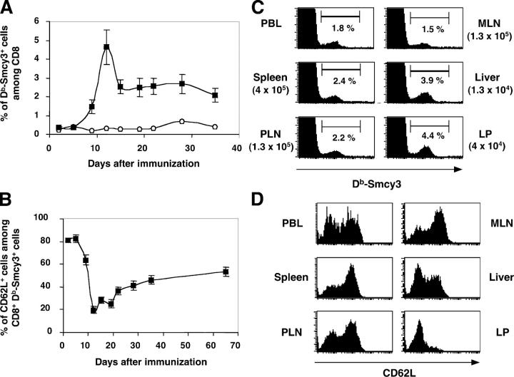
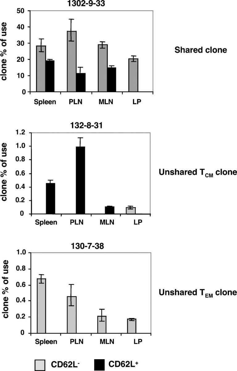
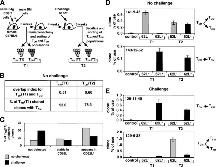
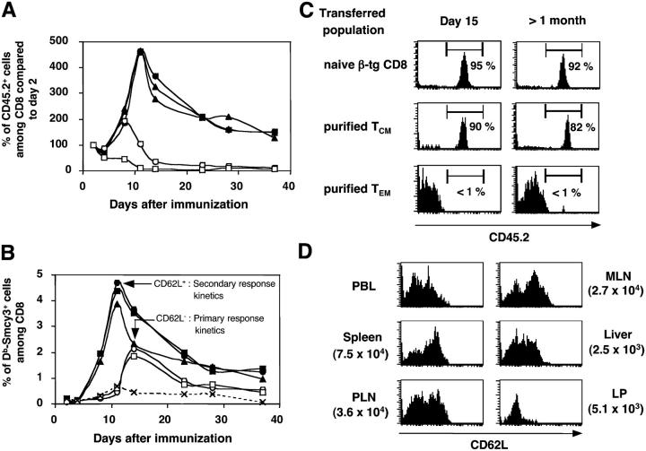
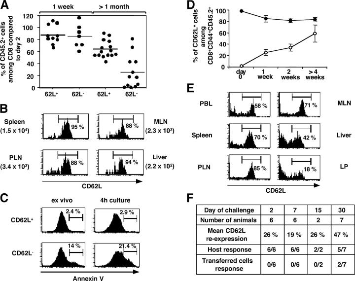
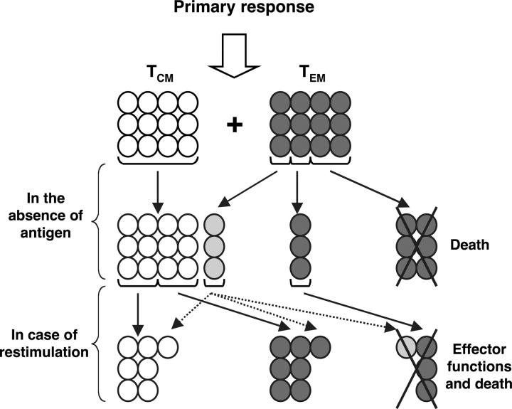
The lineage relationships of central-memory T cells (T(CM)) cells and effector-memory T cells (T(EM)), as well as their homeostasis and recall capacities, are still controversial. We investigated these issues in a murine model using two complementary approaches: T cell receptor repertoire analysis and adoptive transfer experiments of purified H-Y-specific T(CM) and T(EM) populations. Repertoire studies showed that approximately two thirds of T(CM) and T(EM) clones derived from a common naive precursor, whereas the other third was distinct. Both approaches highlighted that T(CM) and T(EM) had drastically distinct behaviors in vivo, both in the absence of antigen or upon restimulation. T(CM) clones were stable in the absence of restimulation and mounted a potent and sustained recall response upon secondary challenge, giving rise to both T(CM) and T(EM), although only a fraction of T(CM) generated T(EM). In contrast, T(EM) persisted for only a short time in the absence of antigen and, although a fraction of them were able to express CD62L, they were unable to mount a proliferative response upon secondary challenge in this model.


Figures

Figure 1.
Figure 1.
In vivo generation of T CD8 memory subsets after intravenous immunization with male bone marrow cells of a C57BL/6 female host transferred with naive β-tg CD8 cells. (A) Analysis of the percentage of Db-Smcy3+ cells among CD8 PBLs for a naive C57BL/6 mouse (open circles) or immunized hosts (closed squares). Data from 12 mice were pooled. (B) Evolution of the percentage of CD62L+ cells among CD8+ Db-Smcy3+ PBLs. (C) Analysis of the percentages and absolute numbers (in brackets) of Db-Smcy3–specific cells among CD8+ cells in several organs 6 wk after immunization. (D) Expression of CD62L by CD8+ Db-Smcy3+ cells in several organs at least 6 wk after immunization. FACS profiles are representative of all mice analyzed.
Figure 2.
Figure 2.
Frequency of individual clones in lymphoid and peripheral organs of an immune mouse. The frequency of each clone among TCRAV9+ cells was measured by quantitative clonotypic PCR within each subset. An example of each type of clone (shared, unshared TCM, unshared TEM) is displayed. The frequency observed in the LP CD62L subset for clone 132–8-31 corresponds to several cells <10 and, therefore, is considered as nonspecific amplification.
Figure 3.
Figure 3.
TCRα repertoire analysis of splenic Db-Smcy3–specific TCM and TEM subsets at two time points (T1 and T2), with or without a challenge in between. TCRAV9-TCRAC rearrangements were sequenced and analyzed. (A) Illustration of the experimental protocol. (B) In the nonchallenged mouse, evolution of the overlap index and of the percentage of shared clones between TEM (T1) and TCM at T1 and T2. (C) Evolution of TEM (T1) unshared clones at T2 in both conditions. (D and E) Evolution of the frequency of T1 unshared clones (D) in the absence of restimulation or (E) in the presence of an intravenous challenge. Frequencies were determined as in Fig. 2.
Figure 4.
Figure 4.
TCM but not TEM can mount a recall response after transfer and challenge. CD62L+ and CD62L cells were purified from the spleen and PLN of hosts transferred with naive β-tg CD8 cells and immunized at least 6 wk earlier (see Materials and methods) and equal numbers of Db-Smcy3–specific TCM (closed symbols) or TEM (open symbols) were transferred into naive CD45.1+ female hosts that were immediately immunized with syngeneic CD45.1+ male bone marrow cells. (A) Evolution of the percentage of CD45.2+ transferred cells among host blood CD8 cells. The percentage observed at day 2 is normalized as 100%, and all subsequent percentages are expressed as a percentage of this standard. (B) Analysis of the percentage of Db-Smcy3+ cells among blood CD8 cells. (C) Expression of the CD45.2 molecule by CD8+ Db-Smcy3+ blood cells at day 15 or >1 mo after challenge, after transfer of naive, TCM, or TEM. (D) CD62L expression profiles and absolute numbers (in parentheses) of CD8+ Db-Smcy3+ cells in organs of a CD45.1+ female host transferred with TCM and challenged at least 6 wk before analysis. All results shown are representative of at least three experiments.
Figure 5.
Figure 5.
Adoptive transfer into naive hosts of CD62L+/− subsets purified from immune mice: overall TCM stability versus TEM death and partial CD62L reexpression. (A, B, and D) TCM and TEM were purified from spleen and PLN of immune hosts and transferred into naive female CD45.1 hosts. (A) Analysis of the persistence of CD8+CD62L+/− transferred cells 1 wk or >1 mo after transfer. The percentage of the CD45.2+ transferred cells among CD8 compared with day 2 is displayed. Each circle represents an individual transferred host. The mean value is shown for each transferred population. (B) CD62L expression and absolute numbers (in parentheses) of CD8+ Db-Smcy3+CD45.2+ cells 6 wk after transfer of purified CD62L+ cells. (C) Analysis of the propensity of TCM and TEM to undergo apoptosis. CD62L+/− subsets were purified from the spleen and PLN cells from an old C57BL/6 mouse, and the expression of annexin V by CD8+CD44+ cells was analyzed either directly ex vivo or after a 4-h stimulation of both subsets with PMA and ionomycin. (D) Kinetic analysis of the percentage of CD62L+ cells among CD8+CD44+CD45.2+ cells in blood after transfer of CD45.2+CD62L+ (closed circles) or CD45.2+CD62L (open circles) cells in a naive CD45.1 host. (E) CD62L expression profiles of CD8+CD45.2+ remaining cells in various organs of a CD45.1 female host 6 wk after transfer of CD62L spleen and PLN cells sorted from an immune donor. (F) Naive CD45.1 female mice were transferred with CD45.2+CD62L cells, allowed to rest for various durations, and challenged with 5 × 10⁶ CD45.1+ male bone marrow cells. Indicated for each duration are the number of mice analyzed, the percentage of transferred CD8 cells expressing CD62L, and the number of individual mice in which a host or a donor response was observed, respectively.
Figure 6.
Figure 6.
Proposed model for the differentiation pathways of TCM and TEM after a primary response, in the absence of antigen, or in the case of antigenic restimulation. TCM are depicted as white cells and TEM are depicted as dark gray cells. The three possible fates of the latter are displayed. Light gray cells represent transitory TEM→TCM reexpressing CD62L but with various recall capacities depending on their position in the TEM→TCM differentiation pathway.

References

    1. Ahmed, R., and D. Gray. 1996. Immunological memory and protective immunity: understanding their relation. Science. 272:54–60. - PubMed
    1. McHeyzer-Williams, M.G., and R. Ahmed. 1999. B cell memory and the long-lived plasma cell. Curr. Opin. Immunol. 11:172–179. - PubMed
    1. Sprent, J., and C.D. Surh. 2002. T cell memory. Annu. Rev. Immunol. 20:551–579. - PubMed
    1. Woodland, D.L., and R.W. Dutton. 2003. Heterogeneity of CD4(+) and CD8(+) T cells. Curr. Opin. Immunol. 15:336–342. - PubMed
    1. Lefrancois, L., and D. Masopust. 2002. T cell immunity in lymphoid and non-lymphoid tissues. Curr. Opin. Immunol. 14:503–508. - PubMed

Publication types

Substances