. 2005 Feb 21;201(4):545-54.
doi: 10.1084/jem.20042060. Epub 2005 Feb 14.

Early appearance of germinal center-derived memory B cells and plasma cells in blood after primary immunization

Affiliations

Early appearance of germinal center-derived memory B cells and plasma cells in blood after primary immunization

Elizabeth J Blink et al. J Exp Med. .

Abstract

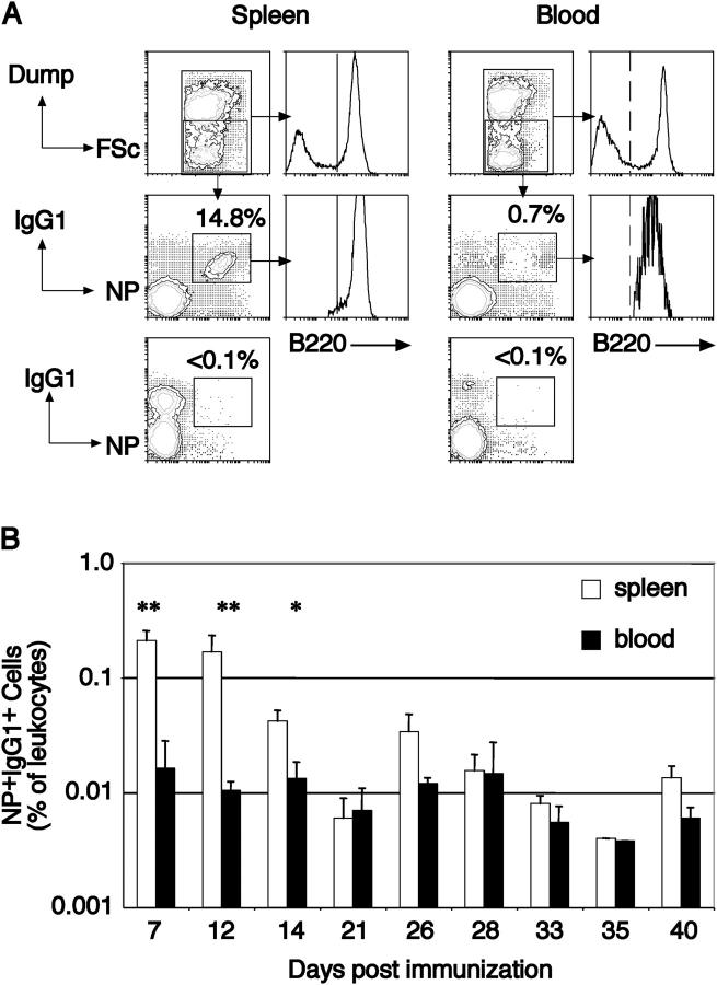
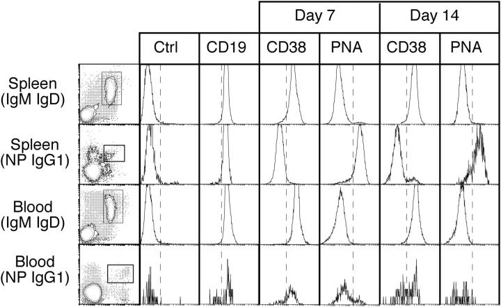
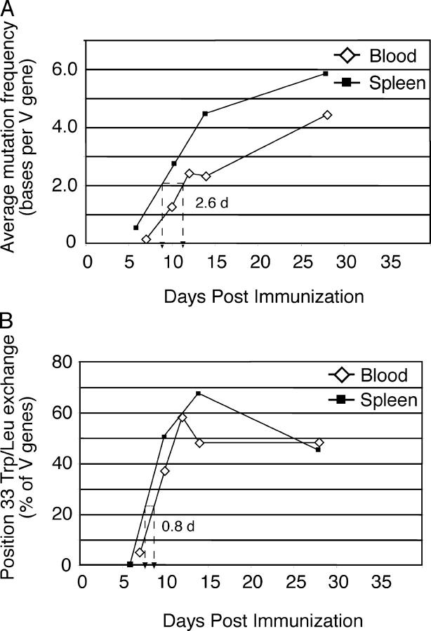
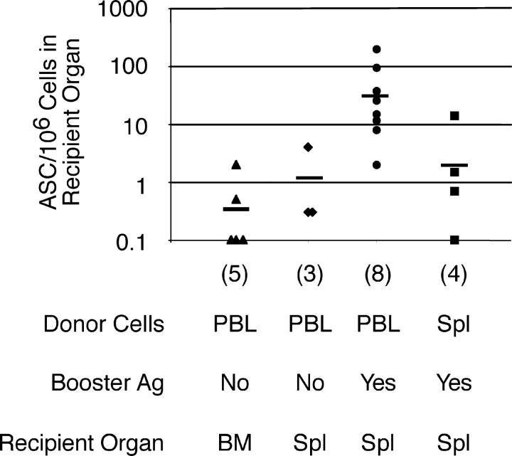
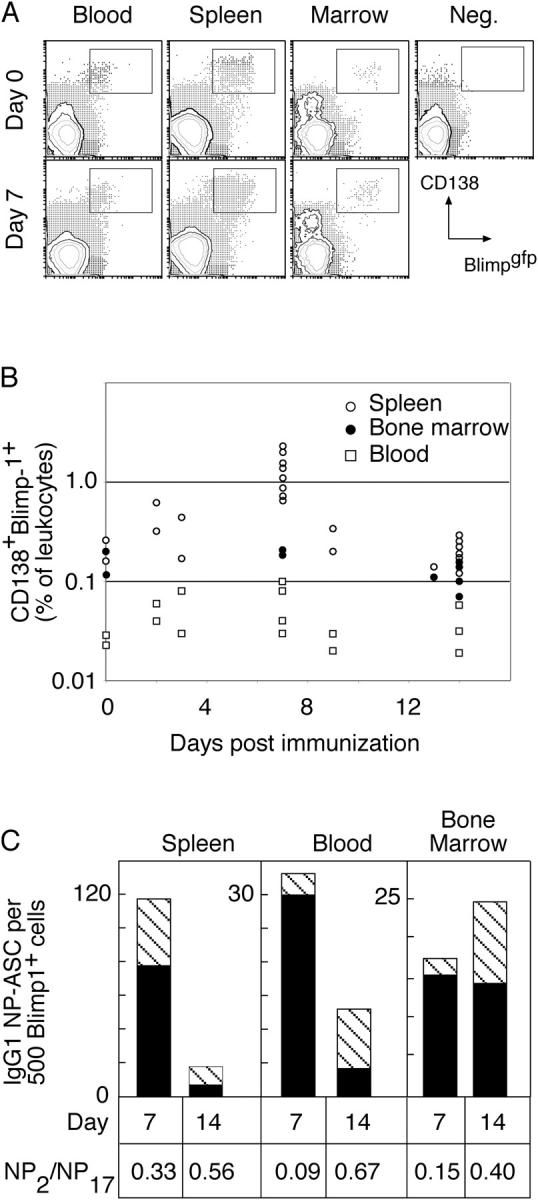
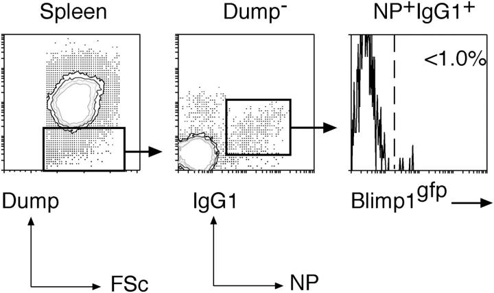
Immunization with a T cell-dependent antigen elicits production of specific memory B cells and antibody-secreting cells (ASCs). The kinetic and developmental relationships between these populations and the phenotypic forms they and their precursors may take remain unclear. Therefore, we examined the early stages of a primary immune response, focusing on the appearance of antigen-specific B cells in blood. Within 1 wk, antigen-specific B cells appear in the blood with either a memory phenotype or as immunoglobulin (Ig)G1 ASCs expressing blimp-1. The memory cells have mutated V(H) genes; respond to the chemokine CXCL13 but not CXCL12, suggesting recirculation to secondary lymphoid organs; uniformly express B220; show limited differentiation potential unless stimulated by antigen; and develop independently of blimp-1 expression. The antigen-specific IgG1 ASCs in blood show affinity maturation paralleling that of bone marrow ASCs, raising the possibility that this compartment is established directly by blood-borne ASCs. We find no evidence for a blimp-1-expressing preplasma memory compartment, suggesting germinal center output is restricted to ASCs and B220(+) memory B cells, and this is sufficient to account for the process of affinity maturation.


Figures

Figure 1.
Figure 1.
Identification of antigen-specific B cells in blood. (A) Leukocytes recovered from the spleen and blood of mice immunized 7 d previously were partitioned using flow cytometry into those expressing and not expressing (boxed) a collection of markers, referred to as the Dump-channel: IgM, IgD, Gr-1, and F4/80. Among negative cells, those that had switched isotype to IgG1 and were capable of binding the immunizing hapten were detected by IgG1-specific anti-sera and NP coupled to the fluorescent protein allophycocyanin, revealing a predominant IgG1+ NP-binding population (boxed). Examining the IgG1+NP+ cells in both spleen and blood revealed these cells to be essentially all B220+, shown in comparison to the B220 distribution on all leukocytes (histograms). Mice immunized with KLH in alum do not develop IgG1+NP+ cells in either blood or spleen (lower contours, already gated as Dump). (B) The kinetics of B220+IgG1+ NP-binding B cells in blood and spleen after primary immunization. Blood and spleen from individual mice were collected at the indicated intervals after a single i.p. immunization and the proportion of B220+ NP-binding IgG1+ cells determined by flow cytometry as depicted in A. The plot shows the average of each tissue with the standard deviation. n ≥ 3 mice for each time point. Significant differences as calculated by Student's t test are indicated. *, P < 0.01; **, P < 0.001.
Figure 2.
Figure 2.
The cell surface phenotype of B220+ NP-binding IgG1+ cells in blood and spleen 7 and 14 d after immunization. B220+ cells were purified mechanically from spleen and blood and stained for surface IgM, IgD, IgG1, and NP. The stains of spleen and blood (IgM, IgD)+ B cells shown in the left column depict before separation, whereas those of NP+IgG1+ B cells depict after separation. Boxed areas indicate the cells analyzed for the parameters depicted by the histograms. The fourth parameter in these analyses is indicated above each column. The dashed line provides a point of reference for comparing samples. The cells were recovered from tissues pooled from two to four mice, and these data are representative of three separate experiments. Ctrl, isotype control antibody.
Figure 3.
Figure 3.
Evidence for somatic mutation and affinity maturation in blood NP+IgG1+ B cells. Single NP-binding IgG1+ B cells were isolated from blood and spleen at the indicated times (for details, see Table I). Analysis of those VH-Cγ1 rearrangements using VH186.2 provided the values depicted in this figure. (A) The average mutation frequency of VH genes at the indicated times is plotted against the time after immunization. The dashed line indicates the time difference between the two populations required to reach an average of 2.0 mutations per VH gene. Accumulation of mutations in blood cells is delayed by ∼2.5 d compared with spleen. (B) The proportion of VH sequences in blood and spleen NP-specific IgG1+ B cells containing a position 33 Y → L exchange as a function of time. Unlike the mutation frequency, cells bearing affinity-enhancing mutations are almost as common in blood as in spleen at each time point. In both plots, spleen values use square symbols, whereas blood uses diamonds. Cells were recovered from tissues pooled from ≥6 mice at each time point.
Figure 4.
Figure 4.
Blood leukocytes from immunized mice respond to antigen in the presence of T cell help. Leukocytes were purified from the pooled blood or spleen of six mice immunized 14 d previously with NP-KLH. 107 of these cells were transferred to mice previously primed with a single injection of HSA in alum. Recipients were either left untreated or boosted with NP-HSA as indicated. 4 d after transfer, recipients were killed and the frequency of NP-specific IgG1 ASCs determined in bone marrow or spleen are indicated. The horizontal bar shows the average for each condition. The resolution of this assay was 0.1 IgG1 ASCs per 106 plated cells. The number of recipients assessed for each condition is indicated. No NP- specific IgG1 ASCs were detected in either the absence of transferred cells or after transfer of leukocytes from naive mice (not depicted).
Figure 5.
Figure 5.
ASCs in blood after immunization revealed by expression of blimp-1. (A) Mice with GFP targeted to one allele of the blimp-1 locus were immunized, and blood, spleen, and bone marrow were collected at the indicated times. Flow cytometric analysis of leukocytes revealed populations of GFP+CD138+ cells in all tissues. (B) The frequency of such cells in individual mice is indicated as a percentage of total blood leukocytes for the different tissues. Data from spleen and bone marrow have been reported previously (reference 31) and are shown here for comparative purposes only. (C) GFP+CD138+ cells were purified by sorting and assessed for the frequency of NP-binding IgG1 ASCs, comparing spleen, blood, and bone marrow populations from the same times. In each case, 500 sort-purified cells were placed in ELISPOT wells coated with either high or low haptenated proteins to detect total or high affinity NP-specific IgG1 ASCs, respectively. The number of spots of each type is plotted and represents the average compiled from four experiments at day 14 and three at day 7, each experiment using tissue pooled from two to four mice. The number of total IgG1 anti-NP ASCs per 500 sorted cells is indicated by the height of the column and the number of high affinity ASCs by the striped section of each column. Unimmunized mice (n = 3) showed no NP- specific IgG1 ASCs in any location. The proportion of total ASCs with high affinity is calculated as NP2/NP17 ASCs. The change in ratio between days 7 and 14 within each population is significantly different (P < 0.05, Student's t test) although the differences between populations at each time are not significantly different.
Figure 6.
Figure 6.
CD138-NP+IgG1+ cells appearing in spleen do not express blimp-1. blimp-1 gfp/+ heterozygous mice were immunized with NP-KLH in alum and 14 d later examined for the appearance in the spleen of NP- specific IgG1+ B cells that lacked expression of CD138 in addition to other Dump-channel markers (IgM, IgD, Gr-1, F4/80). However, among these NP+IgG1+ cells, none were found to express blimp-1 as indicated by the absence of GFP+ cells in the gate. One out of two representative experiments is shown.

Comment in

References

    1. Zinkernagel, R.M. 2000. What is missing in immunology to understand immunity? Nat. Immunol. 1:181–185. - PubMed
    1. Rajewsky, K. 1996. Clonal selection and learning in the antibody system. Nature. 381:751–758. - PubMed
    1. Klein, U., K. Rajewsky, and R. Kuppers. 1998. Human immunoglobulin (Ig)M+IgD+ peripheral blood B cells expressing the CD27 cell surface antigen carry somatically mutated variable region genes: CD27 as a general marker for somatically mutated (memory) B cells. J. Exp. Med. 188:1679–1689. - PMC - PubMed
    1. Tangye, S.G., Y.J. Liu, G. Aversa, J.H. Phillips, and J.E. de Vries. 1998. Identification of functional human splenic memory B cells by expression of CD148 and CD27. J. Exp. Med. 188:1691–1703. - PMC - PubMed
    1. Baine, Y., and G.J. Thorbecke. 1982. Induction and persistence of local B cell memory in mice. J. Immunol. 128:639–643. - PubMed

Publication types

MeSH terms