Comparative Study
. 2005 Apr;144(7):961-71.
doi: 10.1038/sj.bjp.0706122.

Neuritic regeneration and synaptic reconstruction induced by withanolide A

Affiliations
Comparative Study

Neuritic regeneration and synaptic reconstruction induced by withanolide A

Tomoharu Kuboyama et al. Br J Pharmacol. 2005 Apr.

Abstract

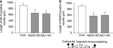
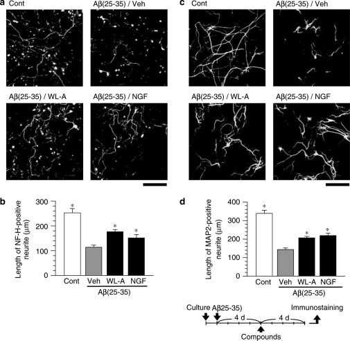
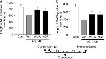
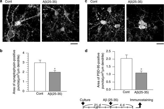
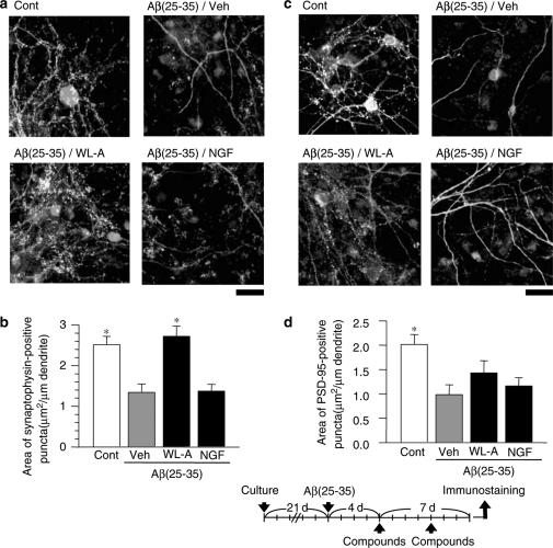
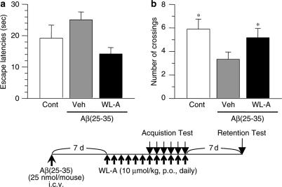
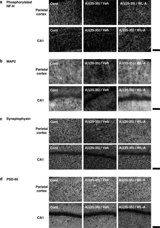
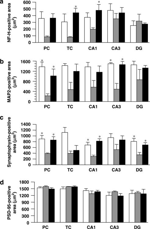
We investigated whether withanolide A (WL-A), isolated from the Indian herbal drug Ashwagandha (root of Withania somnifera), could regenerate neurites and reconstruct synapses in severely damaged neurons. We also investigated the effect of WL-A on memory-deficient mice showing neuronal atrophy and synaptic loss in the brain. Axons, dendrites, presynapses, and postsynapses were visualized by immunostaining for phosphorylated neurofilament-H (NF-H), microtubule-associated protein 2 (MAP2), synaptophysin, and postsynaptic density-95 (PSD-95), respectively. Treatment with A beta(25-35) (10 microM) induced axonal and dendritic atrophy, and pre- and postsynaptic loss in cultured rat cortical neurons. Subsequent treatment with WL-A (1 microM) induced significant regeneration of both axons and dendrites, in addition to the reconstruction of pre- and postsynapses in the neurons. WL-A (10 micromol kg(-1) day(-1), for 13 days, p.o.) recovered A beta(25-35)-induced memory deficit in mice. At that time, the decline of axons, dendrites, and synapses in the cerebral cortex and hippocampus was almost recovered. WL-A is therefore an important candidate for the therapeutic treatment of neurodegenerative diseases, as it is able to reconstruct neuronal networks.


Figures

Figure 1
Figure 1
Axonal and dendritic atrophy induced by Aβ(25–35) and Aβ(1–42). Cortical neurons were cultured for 24 h, and then the cells were treated with 10 μM Aβ(25–35), 10 μM Aβ(1–42), or the vehicle (Cont). At 4 days after treatment, the cells were fixed and immunostained for phosphorylated NF-H or MAP2. Lengths of NF-H-positive (a) or MAP2-positive (b) neurites per cell were measured in each treatment. The values represent the means and s.e.m. of four images. *P<0.05 when compared with Cont.
Figure 2
Figure 2
The effects of withanolide A (WL-A) on axonal and dendritic regeneration after Aβ(25–35)-induced atrophy. Cortical neurons were cultured for 24 h, and were then treated with or without (Cont) 10 μM Aβ(25–35). At 4 days after the administration of Aβ(25–35), the cells were treated with WL-A at a concentration of 1 μM; or NGF at a concentration of 100 ng ml−1; or the vehicle (Veh). At 4 days after treatment, the cells were fixed and immunostained for phosphorylated NF-H (a) or MAP2 (c). The lengths of NF-H-positive (b) or MAP2-positive (d) neurites were measured in each treatment. The values represent the means and s.e.m. of 30 cells. *P<0.05 when compared with Veh. Scale=100 μm.
Figure 3
Figure 3
The effects of withanolide A (WL-A) on axonal and dendritic regeneration after Aβ(1–42)-induced atrophy. Cortical neurons were cultured for 24 h, and were then treated with or without (Cont) 10 μM Aβ(1–42). At 4 days after the administration of Aβ(1–42), the cells were treated with WL-A at a concentration of 1 μM; or NGF at a concentration of 100 ng ml−1; or the vehicle (Veh). At 4 days after treatment, the cells were fixed and immunostained for phosphorylated NF-H or MAP2. The lengths of NF-H-positive (a) or MAP2-positive (b) neurites per cell were measured in each treatment. The values represent the means and s.e.m. of four images. *P<0.05 when compared with Veh.
Figure 4
Figure 4
Pre- and postsynaptic loss induced by Aβ(25–35). After culture for 21 days, the cortical neurons were treated with or without (Cont) 10 μM Aβ(25–35) for 4 days. The cells were then double-immunostained for synaptophysin (a, green color in online version) or PSD-95 (b, green color in online version), plus MAP2 (red color in online version). Areas of synaptophysin- (b) or PSD-95- (d) positive puncta per 1 μm of dendrite were measured. The values represent the means and s.e.m. of 24–28 dendrites. *P<0.05 when compared with Cont. Scale=50 μm.
Figure 5
Figure 5
The effects of withanolide A (WL-A) on pre- and postsynaptic reconstruction after Aβ(25–35)-induced synaptic loss. After culture for 21 days, the cortical neurons were treated with or without (Cont) Aβ(25–35). The cells were then treated with WL-A at a concentration of 1 μM; or NGF at a concentration of 100 ng ml−1; or with the vehicle (Veh). At 7 days after treatment, the cells were double-immunostained for synaptophysin (a, green in online version) or PSD-95 (c, green in online version), plus MAP2 (red in online version). Areas of synaptophysin- (b) PSD-95- (d) positive puncta per 1 μm of dendrites were measured. The values represent the means and s.e.m. of 20–35 dendrites. *P<0.05 when compared with Veh. Scale=20 μm.
Figure 6
Figure 6
The effect of withanolide A on spatial memory deficit induced by Aβ(25–35). (a) Escape latencies of four trials are shown on training day 5 in a Morris water maze. (b) Crossing numbers over the position where the platform had been located were measured for 60 s 7 days after the last training day. This was also 7 days after the discontinuance of drug treatment. The time schedule of the experiment is shown below the figure. The values represent the means and s.e.m. of 6–9 mice. *P<0.05 when compared with Veh.
Figure 7
Figure 7
Effects of withanolide A on neuritic atrophy and synaptic loss in vivo induced by Aβ(25–35). At 2 days after the retention test, the mice were decapitated, and their brains were coronally sliced. The slices were immunostained with phosphorylated NF-H (a), MAP2 (b), synaptophysin (c), and PSD-95 (d). Representative images of brain slices from each group are shown from the parietal cortex (upper) and the hippocampal CA1 (below). Scale=100 μm.
Figure 8
Figure 8
Quantified effects of withanolide A (WL-A) on neuritic atrophy and synaptic loss in vivo induced by Aβ(25–35). NF-H- (a), MAP2- (b), synaptophysin- (c), and PSD-95- (d) positive areas were measured in the parietal cortex (PC) and temporal cortex (TC), and the hippocampal CA1, CA3, and dentate gyrus (DG). Control mice (open columns) were treated with an i.c.v. injection of saline and p.o. administration of the vehicle. Aβ(25–35) i.c.v.-injected mice were treated with p.o. administration of the vehicle (gray columns) or WL-A (black columns). The values represent the means and s.e.m. of three mice. *P<0.05 when compared with Aβ(25–35) plus the vehicle-treated group.

References

    1. AKAMA K.T., MCEWEN B.S. Estrogen stimulates postsynaptic density-95 rapid protein synthesis via the Akt/Protein kinase B pathway. J. Neurosci. 2003;23:2333–2339. - PMC - PubMed
    1. AUDESIRK T., CABELL L., KERN M., AUDESIRK G. Estradiol influences differentiation of hippocampal neurons in vitro through an estrogen receptor-mediated process. Neuroscience. 2003;121:927–934. - PubMed
    1. BOBINSKI M., WEGIEL J., TARNAWSKI M., BOBINSKI M., REISBERG B., DE LEON M.J., MILLER D.C., WISNIEWSKI H.M. Relationships between regional neuronal loss and neurofibrillary changes in the hippocampal formation and duration and severity of Alzheimer disease. J. Neuropath. Exp. Neurol. 1997;56:414–420. - PubMed
    1. CAMBIASSO M.J., CARRER H.F. Nongenomic mechanism mediates estradiol stimulation of axon growth in male rat hypothalamic neurons in vitro. J. Neurosci. 2001;66:475–481. - PubMed
    1. CANNING D.R., MC KEON R.J., DE WITT D.A., PERRY G., WUJEK J.R., FREDERICKSON R.C., SILVER J. Amyloid of Alzheimer's disease induces reactive gliosis that inhibits axonal outgrowth. Exp. Neurol. 1993;124:289–298. - PubMed

Publication types