. 2005 Mar;115(3):599-609.
doi: 10.1172/JCI22304.

Targeted deletion or pharmacological inhibition of MMP-2 prevents cardiac rupture after myocardial infarction in mice

Affiliations

Targeted deletion or pharmacological inhibition of MMP-2 prevents cardiac rupture after myocardial infarction in mice

Shin-ichiro Matsumura et al. J Clin Invest. 2005 Mar.

Abstract

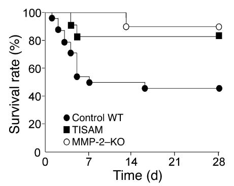
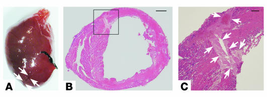
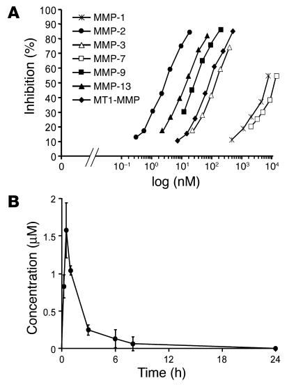
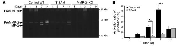
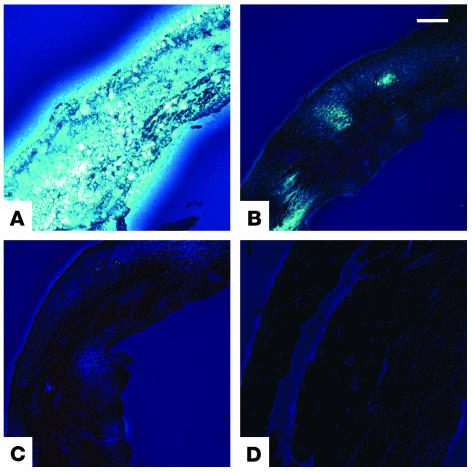
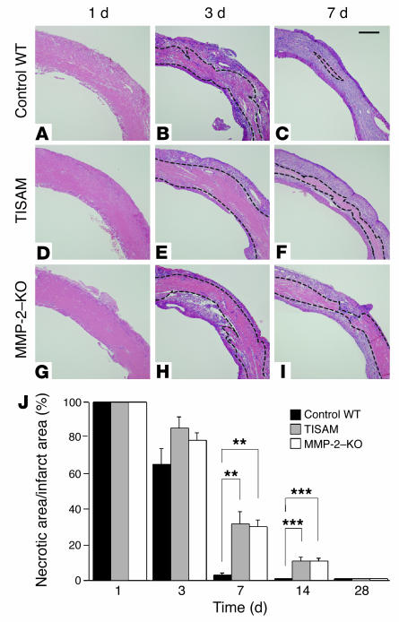
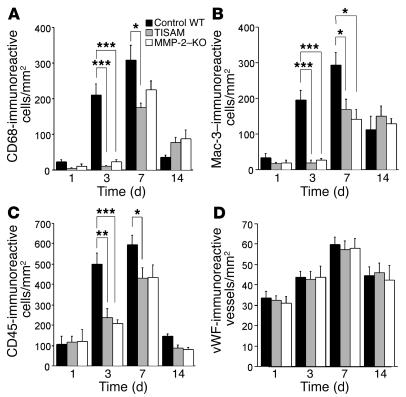
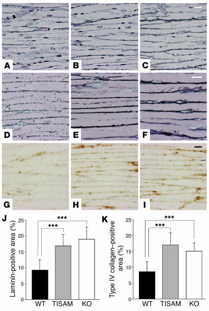
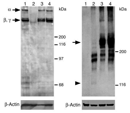
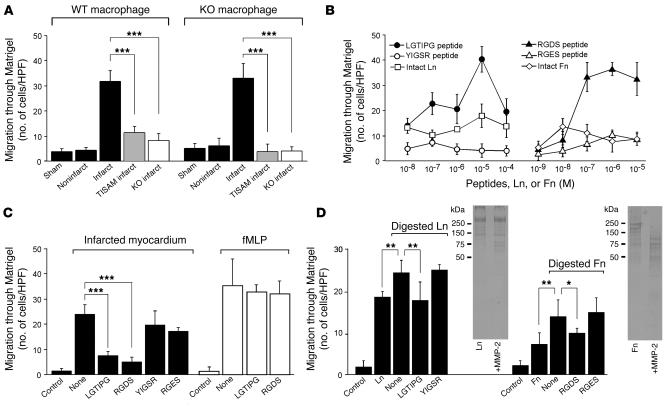
MMPs are implicated in LV remodeling after acute myocardial infarction (MI). To analyze the role of MMP-2, we generated MI by ligating the left coronary artery of MMP-2-KO and WT mice, the latter of which were administered orally an MMP-2-selective inhibitor or vehicle (TISAM). The survival rate was significantly higher in MMP-2-KO and TISAM-treated mice than in control WT mice. The main cause of mortality in control WT mice was cardiac rupture, which was not observed in MMP-2-KO or TISAM-treated mice. Control WT mice, but not MMP-2-KO or TISAM-treated mice, showed activation of the zymogen of MMP-2, strong gelatinolytic activity, and degradation of ECM components, including laminin and fibronectin, in the infarcted myocardium. Although infarcted cardiomyocytes in control WT mice were rapidly removed by macrophages, the removal was suppressed in MMP-2-KO and TISAM-treated mice. Macrophage migration was induced by the infarcted myocardial tissue from control WT mice and was inhibited by treatment of macrophages with laminin or fibronectin peptides prior to migration assay. These data suggest that inhibition of MMP-2 activity improves the survival rate after acute MI by preventing cardiac rupture and delays post-MI remodeling through a reduction in macrophage infiltration.


Figures

Figure 1
Figure 1
Survival of the WT, TISAM-treated, and MMP-2–KO mice after MI. Lifespan was estimated by the Kaplan-Meier method. Percentages of surviving vehicle-treated WT mice (control WT; n = 24), TISAM-treated WT mice (TISAM; n = 11), and MMP-2–KO mice (MMP-2–KO; n = 10) are shown. At 7 and 28 days after coronary ligation, survival rates of TISAM-treated or MMP-2–KO mice were significantly higher than that of control WT mice (P < 0.001).
Figure 2
Figure 2
Macroscopic and microscopic views of MI with LV myocardial rupture in the dead, vehicle-treated WT mice on day 3 after MI. Note that spontaneous rupture is generated in the LV infarct border near the right ventricle (arrows in A and rectangular area in B). (C) High-power view of the rectangular area in B, showing the rupture (arrows). The black string visible in A is a silk suture used for ligation. Scale bars: 500 μm in B and 100 μm in C.
Figure 3
Figure 3
Concentration-dependent inhibition of TISAM by 7 different MMPs and plasma concentration–time profile in mice. (A) The IC50 values log (nM) for MMP-1, -2, -3, -7, -9, and -13 and MT1-MMP were determined based on a nonlinear regression fit of the concentration-dependent reaction rates using quenched fluorescent peptide substrates. (B) The mean plasma concentrations of TISAM at 0, 0.25, 0.5, 1, 3, 6, 8, and 12 hours after an oral administration were measured.
Figure 4
Figure 4
Gelatin zymography of homogenate supernatants from the LV infarcts of vehicle-treated control WT, TISAM-treated, and MMP-2–KO mice and activation ratio of proMMP-2 in control and TISAM-treated mice. (A) Gelatinolytic activity in the myocardial tissues obtained from vehicle-treated WT, TISAM-treated, and MMP-2–KO mice on days 1, 3, 7, and 14. “S” indicates the myocardial samples obtained from sham-operated WT and MMP-2–KO mice on day 7. ProMMP-2 of 63 kDa, active MMP-2 of 57 kDa, and proMMP-9 of 94 kDa are indicated. (B) Activation ratios of proMMP-2 in infarcted myocardial tissues at 0, 1, 3, 7, and 14 days after coronary ligation. The percentage (active MMP-2 divided by the sum of proMMP-2 and active MMP-2) was measured by densitometric analysis of the activity of each species. **P < 0.01; ***P < 0.001.
Figure 5
Figure 5
Demonstration of gelatinolytic activity in the infarcted myocardium by in situ zymography. Frozen sections were prepared from transverse slices of hearts obtained from vehicle-treated control WT mice (A), TISAM-treated mice sacrificed 6 hours after the last administration of TISAM (B), MMP-2–KO mice (C), and sham-operated WT mice (D) and subjected to in situ zymography as described in Methods. Note the inhibition of gelatinolytic activity in TISAM-treated mouse heart (B) and negligible activity in MMP-2–KO mouse heart (C). Scale bar: 200 μm.
Figure 6
Figure 6
Histology and morphometry of the LV infarcted myocardium from vehicle-treated control WT, TISAM-treated, and MMP-2–KO mice. Paraffin sections of the hearts obtained from vehicle-treated control WT mice (AC), TISAM-treated mice (DF), and MMP-2–KO mice (GI) on days 1, 3, and 7 were stained with H&E. Necrotic zones are marked by dotted lines. Scale bar: 300 μm. (J) Morphometrical analysis of the LV necrotic area to total infarct area, showing that phagocytic removal of necrotic cardiomyocytes by macrophages was significantly reduced in TISAM-treated and MMP-2–KO mice on days 7 and 14. **P < 0.01; ***P < 0.001.
Figure 7
Figure 7
Inflammatory cell accumulation and angiogenesis in boundary areas of the infarcted myocardium evaluated by immunohistochemistry. Frozen sections were immunostained for CD68, Mac-3, and CD45, and we determined accumulation of immunoreactive cells, denoted as cells/mm2, by counting immunoreactive cells in 5 different areas of 0.25 mm2 using NIH Image software as described in Methods. Similarly, paraffin sections were immunostained for vWF and immunoreactive blood vessels with an apparent luminal area were counted. (A and B) Infiltration of CD68- or Mac-3–immunoreactive macrophages in infarcted myocardium from vehicle-treated control WT, TISAM-treated, and MMP-2–KO mice on days 1, 3, 7, and 14. (C) Infiltration of CD45-reactive polymorphonuclear leukocytes in infarcted myocardium from control WT, TISAM-treated, and MMP-2–KO mice on days 1, 3, 7, and 14. (D) Angiogenesis in infarcted myocardium from control WT, TISAM-treated, and MMP-2–KO mice on days 1, 3, 7, and 14. *P < 0.05; **P < 0.01; ***P < 0.001.
Figure 8
Figure 8
Degradation of ECM components in infarcted myocardium on day 3. Representative micrographs of transverse sections of sham-operated noninfarcted LV myocardium (AC) and infarcted myocardium (DI) from vehicle-treated WT mice (A, D, and G), TISAM-treated mice (B, E, and H) and MMP-2–KO mice (C, F, and I) stained by silver impregnation (AF) and immunostained with anti-laminin antibody (GI). Note the marked degradation of ECM components stained by silver impregnation (D) and by immunohistochemistry of laminin (G) around the necrotic cardiomyocytes in control WT mice, which contrasts the findings that these components remained in TISAM-treated mice (E and H) and MMP-2–KO mice (F and I). Infarcted cardiomyocytes (DI) are characterized by disappearance of their nuclei. Scale bars: 20 μm (AC), 10 μm (DF), and 20 μm (GI). Quantitative analysis of laminin-positive (J) or type IV collagen–positive structures (K) in the infarcted areas (%) was performed by computerized morphometry. ***P < 0.001.
Figure 9
Figure 9
Demonstration of the degradation of laminin and fibronectin by immunoblotting. Supernatants of infarcted myocardial tissue homogenates were prepared from vehicle-treated WT mice (lane 2), TISAM-treated mice (lane 3), and MMP-2–KO mice (lane 4), subjected to SDS-PAGE, and immunoblotted for laminin (A) and fibronectin (B) as described in Methods. Sham-operated noninfarcted myocardium (lane 1) was also immunoblotted. Note the marked degradation of laminin and fibronectin in infarcted myocardium from vehicle-treated control WT mice, which was almost completely inhibited in TISAM-treated and MMP-2–KO mice. α, β, and γ chains of laminin and intact fibronectin of 240 kDa are indicated by arrows. The arrowhead denotes a fibronectin fragment of 120 kDa. Immunoblotting for β-actin was used to show the similar amount of sample application to each lane.
Figure 10
Figure 10
Macrophage migration in response to infarcted myocardium (A), laminin and fibronectin peptides (B), inhibition of migration activity to infarcted myocardium by laminin and fibronectin peptides (C), and digestion products of laminin and fibronectin by MMP-2 (D). (A) Migration activity of macrophages derived from WT and MMP-2–KO mice in response to infarcted myocardium from vehicle-treated WT (Infarct), TISAM-treated (TISAM infarct), and MMP-2–KO mice (KO infarct). As for controls, myocardium from sham-operated WT mice (Sham) and from noninfarct areas of infarcted vehicle-treated WT mice (Noninfarct) was used. HPF, high-power field. ***P < 0.001. (B) Migration activity of macrophages from WT mice in response to laminin peptides (LGTIPG and YIGSR) and intact laminin (Ln) and fibronectin peptides (RGDS and RGES) and intact fibronectin (Fn). (C) Inhibition of WT macrophage migration activity to infarcted myocardium or fMLP by laminin and fibronectin peptides. Macrophages were incubated with buffer alone (None), laminin peptides, or fibronectin peptides and then subjected to a migration assay by the addition of infarcted myocardium from WT mice or fMLP to lower chambers. Control, GBSS-BSA alone in lower chambers. ***P < 0.001. (D) Macrophage migration in response to MMP-2 digestion products of laminin and fibronectin. Macrophages were incubated with buffer alone, laminin, or fibronectin peptides and then subjected to a migration assay by the addition of the digestion products to lower chambers. Control, Ln, and Fn represent GBSS-BSA alone, laminin alone, and fibronectin alone in lower chambers, respectively. Digestion patterns of laminin and fibronectin on SDS-PAGE are shown. *P < 0.05; **P < 0.01.

References

    1. Antman, E.M., and Braunwald, E. 2001. Acute myocardial infarction. In Harrison’s principles of internal medicine. E. Braunwald, et al., editors. McGraw-Hill. New York, New York, USA. 1386–1399.
    1. Przyklenk K, Connelly CM, McLaughlin RJ, Kloner RA, Apstein CS. Effect of myocyte necrosis on strength, strain, and stiffness of isolated myocardial strips. Am. Heart J. 1987;114:1349–1359. - PubMed
    1. Factor SM, Robinson TF, Dominitz R, Cho SH. Alterations of the myocardial skeletal framework in acute myocardial infarction with and without ventricular rupture. A preliminary report. Am. J. Cardiovasc. Pathol. 1987;1:91–97. - PubMed
    1. Gao XM, et al. Lower risk of postinfarct rupture in mouse heart overexpressing beta 2-adrenergic receptors: importance of collagen content. J. Cardiovasc. Pharmacol. 2002;40:632–640. - PubMed
    1. Ichihara S, et al. Targeted deletion of angiotensin II type 2 receptor caused cardiac rupture after acute myocardial infarction. Circulation. 2002;106:2244–2249. - PubMed

Publication types

MeSH terms

Substances