The transmembrane oligomers of coronavirus protein E
- PMID: 15713601
- PMCID: PMC1305130
- DOI: 10.1529/biophysj.104.051730
The transmembrane oligomers of coronavirus protein E
Abstract
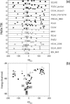
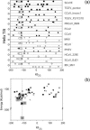
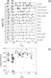
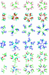
We have tested the hypothesis that severe acute respiratory syndrome (SARS) coronavirus protein E (SCoVE) and its homologs in other coronaviruses associate through their putative transmembrane domain to form homooligomeric alpha-helical bundles in vivo. For this purpose, we have analyzed the results of molecular dynamics simulations where all possible conformational and aggregational space was systematically explored. Two main assumptions were considered; the first is that protein E contains one transmembrane alpha-helical domain, with its N- and C-termini located in opposite faces of the lipid bilayer. The second is that protein E forms the same type of transmembrane oligomer and with identical backbone structure in different coronaviruses. The models arising from the molecular dynamics simulations were tested for evolutionary conservation using 13 coronavirus protein E homologous sequences. It is extremely unlikely that if any of our assumptions were not correct we would find a persistent structure for all the sequences tested. We show that a low energy dimeric, trimeric and two pentameric models appear to be conserved through evolution, and are therefore likely to be present in vivo. In support of this, we have observed only dimeric, trimeric, and pentameric aggregates for the synthetic transmembrane domain of SARS protein E in SDS. The models obtained point to residues essential for protein E oligomerization in the life cycle of the SARS virus, specifically N15. In addition, these results strongly support a general model where transmembrane domains transiently adopt many aggregation states necessary for function.
Figures


Similar articles
-
Model of a putative pore: the pentameric alpha-helical bundle of SARS coronavirus E protein in lipid bilayers.Biophys J. 2006 Aug 1;91(3):938-47. doi: 10.1529/biophysj.105.080119. Epub 2006 May 12. Biophys J. 2006. PMID: 16698774 Free PMC article.
-
Membrane assembly of simple helix homo-oligomers studied via molecular dynamics simulations.Biophys J. 2007 Feb 1;92(3):854-63. doi: 10.1529/biophysj.106.095216. Epub 2006 Nov 3. Biophys J. 2007. PMID: 17085501 Free PMC article.
-
Conductance and amantadine binding of a pore formed by a lysine-flanked transmembrane domain of SARS coronavirus envelope protein.Protein Sci. 2007 Sep;16(9):2065-71. doi: 10.1110/ps.062730007. Protein Sci. 2007. PMID: 17766393 Free PMC article.
-
SARS coronavirus E protein in phospholipid bilayers: an x-ray study.Biophys J. 2006 Mar 15;90(6):2038-50. doi: 10.1529/biophysj.105.072892. Epub 2005 Dec 16. Biophys J. 2006. PMID: 16361349 Free PMC article.
-
How translocons select transmembrane helices.Annu Rev Biophys. 2008;37:23-42. doi: 10.1146/annurev.biophys.37.032807.125904. Annu Rev Biophys. 2008. PMID: 18573071 Review.
Cited by
-
A single polar residue and distinct membrane topologies impact the function of the infectious bronchitis coronavirus E protein.PLoS Pathog. 2012;8(5):e1002674. doi: 10.1371/journal.ppat.1002674. Epub 2012 May 3. PLoS Pathog. 2012. PMID: 22570613 Free PMC article.
-
The transmembrane homotrimer of ADAM 1 in model lipid bilayers.Protein Sci. 2007 Feb;16(2):285-92. doi: 10.1110/ps.062494307. Epub 2006 Dec 22. Protein Sci. 2007. PMID: 17189481 Free PMC article.
-
Structure of a conserved Golgi complex-targeting signal in coronavirus envelope proteins.J Biol Chem. 2014 May 2;289(18):12535-49. doi: 10.1074/jbc.M114.560094. Epub 2014 Mar 25. J Biol Chem. 2014. PMID: 24668816 Free PMC article.
-
Immunoinformatic analysis of the SARS-CoV-2 envelope protein as a strategy to assess cross-protection against COVID-19.Microbes Infect. 2020 May-Jun;22(4-5):182-187. doi: 10.1016/j.micinf.2020.05.013. Epub 2020 May 21. Microbes Infect. 2020. PMID: 32446902 Free PMC article.
-
SARS-CoV-2 Envelope Protein Forms Clustered Pentamers in Lipid Bilayers.Biochemistry. 2022 Nov 1;61(21):2280-2294. doi: 10.1021/acs.biochem.2c00464. Epub 2022 Oct 11. Biochemistry. 2022. PMID: 36219675 Free PMC article.
References
-
- Adams, P. D., I. T. Arkin, D. M. Engelman, and A. T. Brunger. 1995. Computational searching and mutagenesis suggest a structure for the pentameric transmembrane domain of phospholamban. Nat. Struct. Biol. 2:154–162. - PubMed
-
- Arkin, I. T., K. R. MacKenzie, and A. T. Brunger. 1997. Site-directed dichroism as a method for obtaining rotational and orientational constraints for oriented polymers. J. Am. Chem. Soc. 119:8973–8980.
Publication types
MeSH terms
Substances
LinkOut - more resources
Full Text Sources
Miscellaneous

