. 2005 Mar;25(5):1586-95.
doi: 10.1128/MCB.25.5.1586-1595.2005.

RNA polymerase I transcription factors in active yeast rRNA gene promoters enhance UV damage formation and inhibit repair

Affiliations

RNA polymerase I transcription factors in active yeast rRNA gene promoters enhance UV damage formation and inhibit repair

Andreas Meier et al. Mol Cell Biol. 2005 Mar.

Abstract

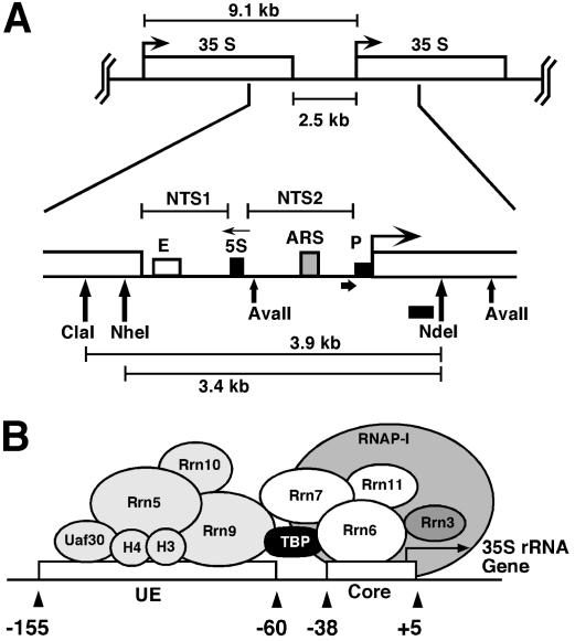
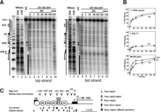
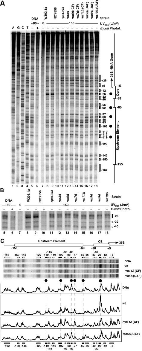
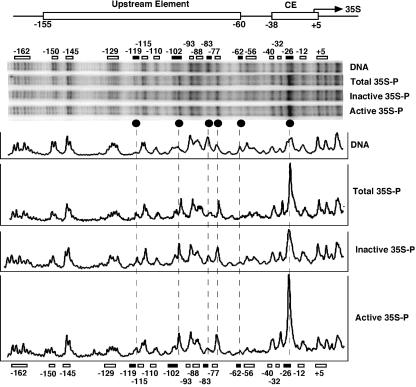
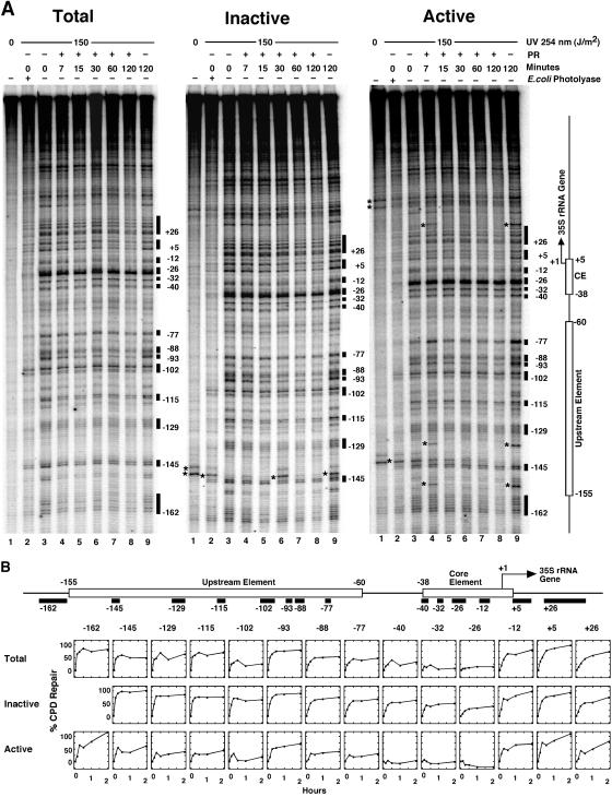
UV photofootprinting and repair of pyrimidine dimers by photolyase was used to investigate chromatin structure, protein-DNA interactions, and DNA repair in the spacer and promoter of Saccharomyces cerevisiae rRNA genes. Saccharomyces cerevisiae contains about 150 copies of rRNA genes separated by nontranscribed spacers. Under exponential growth conditions about half of the genes are transcribed by RNA polymerase I (RNAP-I). Initiation of transcription requires the assembly of the upstream activating factor (UAF), the core factor (CF), TATA binding protein, and RNAP-I with Rrn3p on the upstream element and core promoter. We show that UV irradiation of wild-type cells and transcription factor mutants generates photofootprints in the promoter elements. The core footprint depends on UAF, while the UAF footprint was also detected in absence of the CFs. Fractionation of active and inactive promoters showed the core footprint mainly in the active fraction and similar UAF footprints in both fractions. DNA repair by photolyase was strongly inhibited in active promoters but efficient in inactive promoters. The data suggest that UAF is present in vivo in active and inactive promoters and that recruitment of CF and RNAP-I to active promoters generates a stable complex which inhibits repair.


Figures

FIG. 1.
FIG. 1.
The intergenic rRNA gene spacer of the yeast S. cerevisiae. (A) One unit of the rRNA gene repeat consists of 9.1 kb of DNA. It contains the following elements: the 35S-rRNA gene transcribed by RNAP-I (white box; 35S); a 5S-rRNA gene transcribed by RNAP-III (black box; 5S); a ribosomal origin of replication (gray box; ARS); an enhancer (white box; E); and two nontranscribed spacers between the 35S and 5S genes (NTS1 and NTS2). Relevant restriction sites and fragments (AvaII, ClaI, NheI, NdeI, 3.9 kb, and 3.4 kb), the probe used for indirect end labeling from the NdeI site (black bar), and the primer used for high resolution footprinting (fat horizontal arrow) are indicated. (B) Schematic illustration of the promoter elements and transcription factors: upstream element (UE), core element (core), UAF containing Rrn5, Rrn9, Rrn10, Uaf30, histone H3, and histone H4, the CF containing Rrn6, Rrn7, Rrn11, the TATA binding protein (TBP), and the RNA-polymerase I (RNAP-I) with the associated Rrn3 (36).
FIG. 2.
FIG. 2.
Chromatin structure modulates UV damage formation and repair in the rRNA gene spacer. (A) Chromatin footprinting by micrococcal nuclease (MNase) was compared with CPD repair by photolyase in AMY3 (rad1Δ). Cells were irradiated with UV light (150 J/m2) and exposed to photoreactivating light (PR). DNA was purified, digested with ClaI and NdeI, and cut at CPDs with T4-endoV. Chromatin (CHR) and genomic DNA (DNA) were isolated from UCC510, partially digested with micrococcal nuclease (MNase), and cut with ClaI and NdeI. The DNA was fractionated on alkaline agarose gels and blotted and hybridized with probes for the bottom strand (right panel) and top strand (left panel), respectively. The positions of the DNA elements (as described for Fig. 1A), positioned nucleosomes (white circles), and nucleosomes not positioned (overlapping circles) are indicated. M, a size marker with multiples of 256 bp; lanes 1, DNA digested with MNase; lanes 2, chromatin digested with MNase; lanes 3, DNA of unirradiated cells; lanes 4, DNA of irradiated cells with no repair (initial damage); lanes 5 to 9, DNA of cells after incubation in photoreactivating light for 7 to 120 min; lanes 10, DNA of cells after incubation in the dark for 120 min. Sites of differential repair in the ARS region (dot, triangles) and in the promoter (star) are indicated. (B) Repair curves of the whole spacer region (white bar in panel A), the promoter region (35S-P; star in panel A), and the transcribed region (black bar in panel A). Black symbols, bottom stand; white symbols, top strand. The bottom strand is the transcribed strand (TS); the top strand is the nontranscribed strands (NTS) of the 35S gene. Data are given as averages with standard deviations for at least three gels. The modulation of repair in the spacer, promoter, and coding region was reproduced in duplicate experiments. Strand-specific repair in the coding region confirmed previous observations (30). (C) Schematic summary and structural interpretation of DNA accessibility to MNase (arrowheads) and photolyase (triangles). Circles 1, 3, 4, and 5 represent positions of nucleosomes. Whether ARS is included in a nucleosome is unclear (circled question mark; see text).
FIG. 3.
FIG. 3.
Inhibition of photoreactivation in active promoters. (A) Schematic illustration of five rRNA gene repeats with the 35S rRNA genes (boxes), promoters (triangles), and spacer and restriction sites for NheI (short arrows) and NarI (long arrows). (B) Fractionation procedure for active and inactive 35S promoters in rRNA gene chromatin. White and black triangles and boxes depict active and inactive promoters and genes, respectively. Spacers and inactive genes are packaged in nucleosomes (grey-shaded circles); active genes are free of nucleosomes (11). Panel 1: nuclear chromatin was digested with NheI to release transcriptionally active genes (30, 32) and a fragment containing an rRNA gene spacer flanked by active genes. All other promoters remain in long, uncut chromatin fragments. Panel 2: DNA was purified. Panel 3: DNA was digested with NarI, which generates spacers containing fragments of 4.7, 8.4, 9.1, and 5.5 kb. Panel 4: the DNA was fractionated on an agarose gel, and fragments containing active promoters (open triangles; 4.7 and 5.5 kb) and inactive promoters (black triangles; 8.4 and 9.1 kb) were excised and purified. (C) AMY3cells were irradiated as described for Fig. 2. DNA fragments containing promoters of active and inactive genes were purified (as depicted in panel B), digested with NheI and NdeI, cut at CPDs with T4-endoV (lanes 2 to 8), fractionated on alkaline agarose gels, and blotted and hybridized with strand-specific probes for the bottom strand (see Fig. 1A). Lanes 1, same as lanes 2 but with no T4-endoV treatment; M, size marker as described for Fig. 2. The 35S promoter is marked with a star.
FIG. 4.
FIG. 4.
UV photofootprinting in 35S promoter of yeast strains defective in RNAP-I transcription factors. (A) Yeast strains were irradiated with 150 J/m2 in selective medium containing galactose. DNA was purified and digested with AvaII. An end-labeled primer was annealed to the bottom strand 280 bp upstream of the transcription initiation site (fat horizontal arrow in Fig. 1A) and was extended by use of Taq polymerase towards the transcription start site of the 35S gene. Products were separated on a 5% acrylamide-42% urea sequencing gel. Lanes 1 to 4, dideoxy sequencing reactions. Lane 5, DNA damaged in vitro with 80 J/m2. Lane 6, damaged DNA as described for lane 5treated with E. coli photolyase. Lane 7, DNA of nonirradiated W303.1a cells. Lanes 8 to 18, DNA of different yeast strains irradiated with 150 J/m2 (chromatin). Lane 8, W303.1a. Lane 9 and 10, NOY556. Lane 10, treatment with E. coli photolyase in vitro. Lane 11, NOY408-1a (rpa135Δ; defective in RNAP-I). Lane 12, NOY604 (rrn3Δ). Lane 13 to 15, strains with mutations in the CF: NOY567 (rrn6Δ), NOY558 (rrn7Δ), and NOY730 (rrn11Δ). Lane 16 to 18, strains with mutations in the UAF: NOY699 (rrn5Δ), NOY703 (rrn9Δ), and NOY704 (rrn10Δ). The elements of the promoter region (as described for Fig. 1) and damage clusters and their positions with respect to the transcription initiation site (+1) (black and white boxes) are indicated. (B) A magnification of the core element region. (C) Pyrimidine dimer patterns in the promoter region of irradiated DNA and chromatin of strain NOY556 (wt), the CF mutant NOY730 (rrn11Δ), and the UAF mutant NOY699 (rrn5Δ). A comparison of relative pyrimidine dimer yields is shown by PhosphorImager scanning results. Black circles and dashed lanes depict dimer positions with marked differences in damage formation (UV photofootprint; see text for details).
FIG. 5.
FIG. 5.
UV photofootprints in active and inactive promoters. DNA was irradiated with 80 J/m2. AMY3 (rad1Δ) cells were irradiated with 150 J/m2. Active and inactive 35S promoters were isolated as described for Fig. 3. Pyrimidine dimers were detected by primer extension as described for Fig. 4. Pyrimidine dimer distribution in irradiated naked DNA (DNA), in total DNA of irradiated cells (Total 35S-P), and in purified fragments containing active and inactive promoters (Inactive 35S-P; Active 35S-P) are shown. A comparison of relative pyrimidine dimer yields is shown (PhosphorImager scans). Symbols are as defined for Fig. 4C.
FIG. 6.
FIG. 6.
Modulation of photoreactivation in active and inactive promoters. (A) Irradiation of AMY3 and photoreactivation and fractionation of active and inactive 35S promoters were done as described for Fig. 2 and 3. The 35S promoter was analyzed by primer extension as described for Fig. 4. A set of data are shown for total, inactive, and active 35S promoters. The lanes in each set represent DNA of nonirradiated cells (lanes 1); DNA of cells irradiated with 150 J/m2 (lanes 2 to 9) and photoreactivated for 7 to 120 min (lanes 4 to 8) or incubated in the dark for 120 min (lanes 9); and damaged DNA as described for lane 3 but treated with E. coli photolyase (lanes 2). Damage sites used for quantification of repair and their position with respect to the transcription initiation site (+1) are indicated as black boxes. (B) Repair curves are shown for total 35S promoters (Total) and purified inactive and active 35S promoters (Inactive and Active). Repair curves represent the averages of results obtained with two gels (Total and Inactive) or three gels (Active) of the same repair experiment. Differential repair was confirmed in an independent experiment (data not shown).

Similar articles

Cited by

References

    1. Aboussekhra, A., and F. Thoma. 1999. TATA-binding protein promotes the selective formation of UV-induced (6-4)-photoproducts and modulates DNA repair in the TATA box. EMBO J. 18:433-443. - PMC - PubMed
    1. Aprikian, P., B. Moorefield, and R. H. Reeder. 2001. New model for the yeast RNA polymerase I transcription cycle. Mol. Cell. Biol. 21:4847-4855. - PMC - PubMed
    1. Banditt, M., T. Koller, and J. M. Sogo. 1999. Transcriptional activity and chromatin structure of enhancer-deleted rRNA genes in Saccharomyces cerevisiae. Mol. Cell. Biol. 19:4953-4960. - PMC - PubMed
    1. Becker, M. M., and J. C. Wang. 1984. Use of light for footprinting DNA in vivo. Nature 309:682-687. - PubMed
    1. Bordi, L., F. Cioci, and G. Camilloni. 2001. In vivo binding and hierarchy of assembly of the yeast RNA polymerase I transcription factors. Mol. Biol. Cell 12:753-760. - PMC - PubMed

Publication types

MeSH terms

LinkOut - more resources