. 2005 Mar;25(5):1655-68.
doi: 10.1128/MCB.25.5.1655-1668.2005.

The Saccharomyces cerevisiae YLL012/YEH1, YLR020/YEH2, and TGL1 genes encode a novel family of membrane-anchored lipases that are required for steryl ester hydrolysis

Affiliations

The Saccharomyces cerevisiae YLL012/YEH1, YLR020/YEH2, and TGL1 genes encode a novel family of membrane-anchored lipases that are required for steryl ester hydrolysis

René Köffel et al. Mol Cell Biol. 2005 Mar.

Abstract

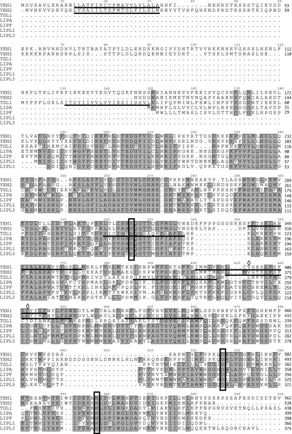
Sterol homeostasis in eukaryotic cells relies on the reciprocal interconversion of free sterols and steryl esters. The formation of steryl esters is well characterized, but the mechanisms that control steryl ester mobilization upon cellular demand are less well understood. We have identified a family of three lipases of Saccharomyces cerevisiae that are required for efficient steryl ester mobilization. These lipases, encoded by YLL012/YEH1, YLR020/YEH2, and TGL1, are paralogues of the mammalian acid lipase family, which is composed of the lysosomal acid lipase, the gastric lipase, and four novel as yet uncharacterized human open reading frames. Lipase triple-mutant yeast cells are completely blocked in steryl ester hydrolysis but do not affect the mobilization of triacylglycerols, indicating that the three lipases are required for steryl ester mobilization in vivo. Lipase single mutants mobilize steryl esters to various degrees, indicating partial functional redundancy of the three gene products. Lipase double-mutant cells in which the third lipase is expressed from the inducible GAL1 promoter have greatly reduced steady-state levels of steryl esters, indicating that overexpression of any of the three lipases is sufficient for steryl ester mobilization in vivo. The three yeast enzymes constitute a novel class of membrane-anchored lipases that differ in topology and subcellular localization.


Figures

FIG. 1.
FIG. 1.
Lack of YEH1 affects the efficiency of steryl ester mobilization in vivo. (A) Wild-type (BY4742, YRS1533) and yeh1Δ (YRS1972), yeh2Δ (YRS1971), and tgl1Δ (YRS1973) mutant cells were labeled for 16 h with [3H]palmitic acid, and the kinetics of steryl ester mobilization in vivo was analyzed by determining steryl ester levels at 0, 2, 4, and 6 h after dilution of the cells in medium containing terbinafine. Lipids were extracted and analyzed by TLC as described in Materials and Methods. TAG, triacylglycerol; STE, steryl esters. (B) The content of [3H]palmitic acid-labeled steryl ester was quantified by radioscanning of TLC plates and set in relation to the levels at time zero (100%). Values represent means and standard deviations of two independent experiments.
FIG. 2.
FIG. 2.
yeh1Δ yeh2Δ tgl1Δ triple-mutant cells fail to mobilize steryl esters in vivo. (A) Lipase triple-mutant (yeh1Δ yeh2Δ tgl1Δ, YRS1840) and double-mutant (yeh1Δ tgl1Δ, YRS1837; yeh2Δ tgl1Δ, YRS1838; yeh1Δ yeh2Δ, YRS1948) cells were labeled for 16 h with [3H]palmitic acid, and the kinetics of steryl ester mobilization in vivo was analyzed after dilution of the cells in medium containing terbinafine. Lipids were extracted and analyzed by TLC as described in Materials and Methods. TAG, triacylglycerol; STE, steryl esters. (B) The content of [3H]palmitic acid-labeled steryl ester was quantified by radioscanning of TLC plates. Values represent means and standard deviations of two independent experiments.
FIG. 3.
FIG. 3.
YEH1, YEH2, and TGL1 are not required for efficient mobilization of triacylglycerol. (A) Wild-type (BY4742, YRS1533) and yeh1Δ (YRS1972), yeh2Δ (YRS1971), and tgl1Δ (YRS1973) mutant cells were labeled for 16 h with [3H]palmitic acid, and the kinetics of triacylglycerol (TAG) mobilization was analyzed after dilution of the cells in medium containing cerulenin. Lipids were extracted and analyzed by TLC, and the relative content of [3H]palmitic acid-labeled TAG was quantified by radioscanning of TLC plates. Values represent means and standard deviations of two independent experiments. (B) Wild-type (BY4742, YRS1533) and lipase triple-mutant (yeh1Δ yeh2Δ tgl1Δ, YRS1840), and double-mutant (yeh1Δ tgl1Δ, YRS1837; yeh2Δ tgl1Δ, YRS1838; yeh1Δ yeh2Δ, YRS1948) cells were labeled for 16 h with [3H]palmitic acid, and the kinetics of TAG mobilization was analyzed as described for panel A.
FIG. 4.
FIG. 4.
Sequence alignment of the three yeast lipases with the mammalian acid lipase family. Sequence alignment of yeast lipases Yeh1 (Q07804), Yeh2 (Q07950), and Tgl1 (P34163) with mammalian acid lipase family members LIPA (P38571), LIPF (P07098), LIPL1 (Q5W064), and LIPL3 (Q5VYY2). Residues of the catalytic triad are boxed. The two functionally important cysteine residues of LIPA are indicated by open diamonds. The positions of potential transmembrane domains in the three yeast lipases, as predicted by TMpred (http://www.ch.embnet.org/software/TMPRED_form.html) and Phobius (http://phobius.cgb.ki.se/), are indicated by horizontal lines. Alignment was performed with ClustalW (http://www.ebi.ac.uk/clustalw/), and regions of homology were drawn by JavaShade (http://industry.ebi.ac.uk/JavaShade/).
FIG. 5.
FIG. 5.
Subcellular localization of Yeh1, Yeh2, and Tgl1. (A) Functionality of the GFP-tagged lipases. Wild-type (YRS1953), triple-mutant (YRS1956), and double-mutant cells in which the remaining lipase is C-terminally tagged with GFP (YRS2090, YRS2091, and YRS2092) were labeled for 16 h with [3H]palmitic acid, and the kinetics of steryl ester mobilization in vivo was analyzed by determining steryl ester levels at 0, 2, 4, and 6 h after dilution of the cells in medium containing terbinafine. Lipids were extracted and analyzed by TLC as described in Materials and Methods. The content of [3H]palmitic acid-labeled steryl ester was quantified by radioscanning of TLC plates and set in relation to the levels at time zero (100%). Values represent means and standard deviations of two independent experiments. (B) In vivo localization of Yeh1-GFP, GFP-Yeh2, and Tgl1-GFP. Heterozygous diploid cells expressing chromosomally tagged versions of Yeh1-GFP and Erg6-RFP (YRS1861) or Tgl1-GFP and Erg6-RFP (YRS2105) were grown in YPD medium and examined by fluorescence microscopy. N-terminally GFP-tagged Yeh2 (YRS2085) was localized after induction in galactose-containing medium for 4 h and stained for DNA with DAPI. Bar, 5 μm. (C) Yeh1, Yeh2, and Tgl1 are membrane associated. Homogenates (Hom) from cells expressing the GFP-tagged lipases were fractionated by differential centrifugation to yield 13,000 × g (13k), 30,000 × g (30k), and 100,000 × g (100k) membrane pellets and cytosolic supernatants (Cyt). Ten-microgram samples of proteins from each fraction were separated by electrophoresis, blotted, and probed with an anti-GFP antibody. (D) Yeh1 is enriched in lipid particles. Haploid cells expressing Yeh1-GFP (YRS1974) were cultivated in rich medium. Vacuoles and lipid particles were isolated by flotation. Homogenate (Hom, 10 μg), vacuole (Vac, 10 μg), and lipid particle (LP, 1 μg) proteins were separated by electrophoresis, blotted, and probed with antibodies against GFP (anti-GFP) and Erg6 (anti-Erg6). The positions of molecular size markers are indicated to the left. (E) Yeh2-GFP cofractionates with plasma membrane markers. Membranes from cells expressing Yeh2-GFP (YRS2086) were fractionated on an Accudenz density gradient, and the presence of Yeh2-GFP was detected by Western blotting with an anti-GFP antibody. The distribution of marker proteins in individual fractions from the gradient was determined by Western blotting with antibodies to Erg6, a marker protein for lipid particles (LP) (8); Pho8-anti-Pho8p, a vacuolar phosphatase (VAC); Pma1, the plasma membrane proton pump (PM); Kar2, an ER luminal chaperone (ER) (15); Mnn1, a medial-Golgi α1,3-mannosyltransferase (GOLGI) (20); Sed5, a t-SNARE of the cis-Golgi (21); and Tlg1, an endosomal-Golgi t-SNARE (GOLGI/EE) (23). (F) Yeh2-GFP is enriched in the plasma membrane fraction. Plasma membrane was enriched by centrifugation of membranes from cells expressing Yeh2-GFP (YRS2086) on a sucrose step gradient and probed for the presence of the GFP epitope, the GPI-anchored plasma membrane protein Gas1, the lipid particle marker Erg6, the ER luminal protein Kar2, and porin, as a marker protein for mitochondria.
FIG. 6.
FIG. 6.
Lipase triple-mutant cells lack steryl ester hydrolase activity in vitro. (A) In vitro steryl ester hydrolase activity in lipase single-, double-, and triple-mutant cells. Wild-type (wt; BY4742, YRS1533) and lipase single (yeh1Δ, YRS1972; yeh2Δ, YRS1971; tgl1Δ, YRS1973)-, double (yeh1Δ tgl1Δ, YRS1837; yeh2Δ tgl1Δ, YRS1838; yeh1Δ yeh2Δ, YRS1948)-, and triple (yeh1Δ yeh2Δ tgl1Δ, YRS1840)-mutant cells were grown in YPD medium and broken with glass beads, and membranes were pelleted. The membrane pellet was solubilized with Triton X-100, and steryl ester hydrolase activity in the detergent extract was determined with cholesterol-[1-14C]oleate as the substrate. The reaction was terminated after 60 min at 30°C, lipids were extracted and separated by TLC, and the released [14C]oleic acid was quantified by radioscanning. The position of the liberated oleic acid is indicated (FFA). The specific activity of steryl ester hydrolase is indicated above the TLC lanes. nd, not detectable. (B) Tgl1-dependent steryl ester hydrolase activity is enriched in lipid particles. Lipid particles from cells of the indicated genotype were isolated, and the steryl ester hydrolase activity was determined in vitro. (C) Yeh2-dependent activity is enriched in the plasma membrane. Plasma membrane from cells of the indicated genotype was isolated, and steryl ester hydrolase activity was determined in vitro.
FIG. 7.
FIG. 7.
Overexpression of N-terminally tagged lipases is sufficient to reduce steryl ester levels. (A) Overexpression of the lipases in a lipase double-mutant background. Strains (YRS2161, YRS2162, and YRS2163) that express an N-terminally GFP-tagged lipase under the control of a galactose-inducible promoter in the absence of the other two lipases were cultivated in either glucose- or galactose-containing rich medium for 16 h. Proteins were extracted, and equal amounts were subjected to Western blot analysis with an antibody against GFP and ER luminal Kar2. (B) Overexpression of GFP-Yeh1, GFP-Yeh2, and GFP-Tgl1 reduces steryl ester but not triacylglycerol levels in vivo. Cells of the indicated genotype were cultivated in either glucose- or galactose-containing rich medium and labeled with [3H]palmitic acid for 16 h. The relative proportions of [3H]palmitic acid in the steryl ester (STE) (B) and triacylglycerol (TAG) (C) pools were quantified by radioscanning of TLC plates. Values represent means and standard deviations of two independent experiments.
FIG. 8.
FIG. 8.
Yeh1, Yeh2, and Tgl1 are integral membrane proteins. (A) Yeh1-, Yeh2-, and Tgl1-GFP are solubilized by detergents. Strains expressing the C-terminally (C-term.) tagged lipases (Yeh1-GFP, YRS1974; Yeh2-GFP, YRS2986; Tgl1-GFP, YRS1858) were grown in YPD medium, cells were broken with glass beads, and membranes were pelleted by centrifugation at 13,000 × g. Fifty micrograms of protein from the membrane pellet was incubated for 30 min at 4°C with 1 M NaCl, 0.1 M Na2CO3, 1% Triton X-100 (TX100), 1% SDS, or buffer alone (Mock) and then centrifuged at 13,000 × g for 30 min to yield soluble (S) and pellet (P) fractions. Proteins were precipitated by TCA, and 10 μg was separated by electrophoresis and probed for the presence of the GFP epitope with an anti-GFP antibody. (B) Protease sensitivity of the C-terminal GFP on Yeh1, Yeh2, and Tgl1. Fifty micrograms of protein from either the 13,000 × g membrane pellet (Yeh1-GFP and Tgl1-GFP) or the enriched plasma membrane fraction (Yeh2-GFP) was incubated with the indicated proteinase K (PrK) concentration for 30 min on ice. Proteins were precipitated by TCA, and 10 μg was separated by electrophoresis and probed for the presence of the GFP epitope or for Kar2 and Gas1, respectively. (C) Protease sensitivity of the N-terminal (N-term.) GFP on Yeh1, Yeh2, and Tgl1. Strains expressing the N-terminally tagged lipases (GFP-Yeh1, YRS2083; GFP-Yeh2, YRS2085; GFP-Tgl1, YRS2084) were grown in YPGal medium, cells were broken with glass beads, and proteins from either the 13,000 × g membrane pellet (GFP-Yeh1 and GFP-Tgl1) or the enriched plasma membrane fraction (GFP-Yeh2) were incubated with the indicated proteinase K concentration for 30 min on ice. Proteins were precipitated by TCA, separated by electrophoresis, and probed for the presence of the GFP epitope and for Kar2 and Gas1, respectively.
FIG. 9.
FIG. 9.
Schematic representation of the proposed membrane topology of yeast lipases Yeh1, Yeh2, and Tgl1 with regard to the catalytic-triad residues.

Similar articles

Cited by

References

    1. Abraham, P. R., A. Mulder, J. Van 't Riet, R. J. Planta, and H. A. Raue. 1992. Molecular cloning and physical analysis of an 8.2 kb segment of chromosome XI of Saccharomyces cerevisiae reveals five tightly linked genes. Yeast 8:227-238. - PubMed
    1. Alani, E., L. Cao, and N. Kleckner. 1987. A method for gene disruption that allows repeated use of URA3 selection in the construction of multiply disrupted yeast strains. Genetics 116:541-545. - PMC - PubMed
    1. Ameis, D., M. Merkel, C. Eckerskorn, and H. Greten. 1994. Purification, characterization and molecular cloning of human hepatic lysosomal acid lipase. Eur. J. Biochem. 219:905-914. - PubMed
    1. Anderson, R. A., R. S. Byrum, P. M. Coates, and G. N. Sando. 1994. Mutations at the lysosomal acid cholesteryl ester hydrolase gene locus in Wolman disease. Proc. Natl. Acad. Sci. USA 91:2718-2722. - PMC - PubMed
    1. Anderson, R. A., and G. N. Sando. 1991. Cloning and expression of cDNA encoding human lysosomal acid lipase/cholesteryl ester hydrolase. Similarities to gastric and lingual lipases. J. Biol. Chem. 266:22479-22484. - PubMed

Publication types

MeSH terms