. 2005 Mar;25(5):1696-712.
doi: 10.1128/MCB.25.5.1696-1712.2005.

Epithelium-mesenchyme interactions control the activity of peroxisome proliferator-activated receptor beta/delta during hair follicle development

Affiliations

Epithelium-mesenchyme interactions control the activity of peroxisome proliferator-activated receptor beta/delta during hair follicle development

Nicolas Di-Poï et al. Mol Cell Biol. 2005 Mar.

Abstract

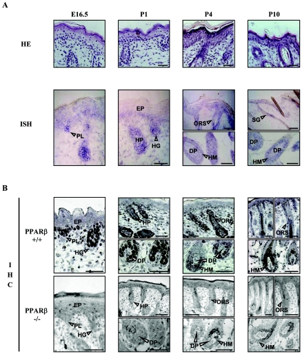
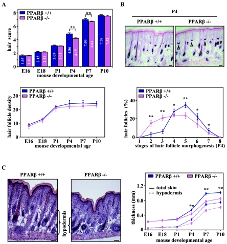
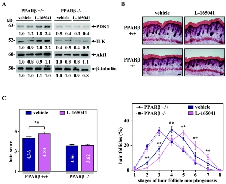
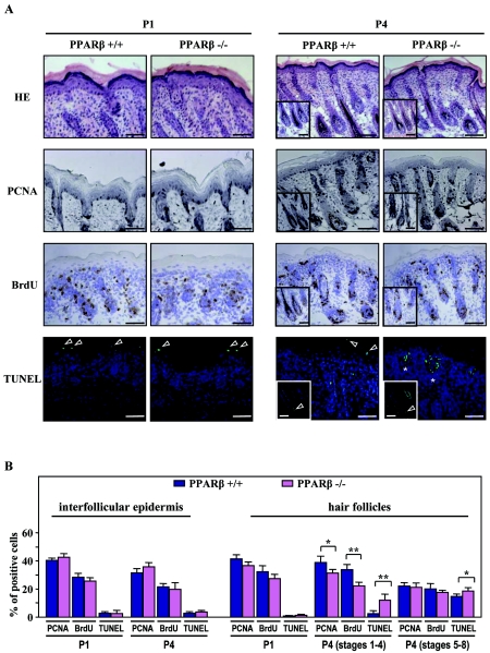
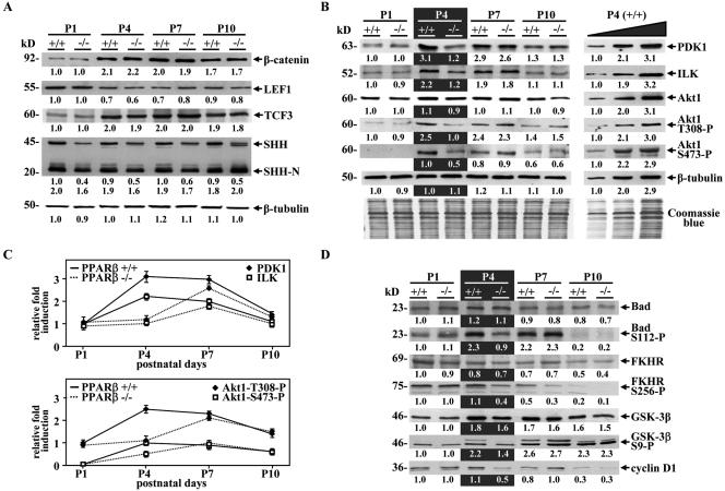
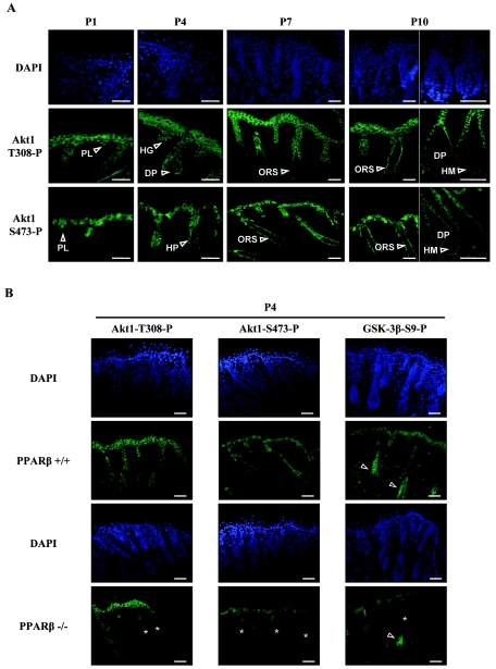
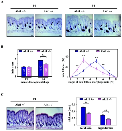
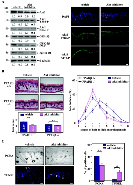
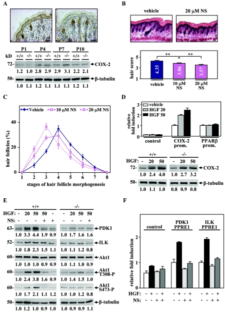
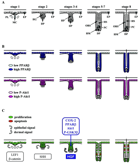
Hair follicle morphogenesis depends on a delicate balance between cell proliferation and apoptosis, which involves epithelium-mesenchyme interactions. We show that peroxisome proliferator-activated receptor beta/delta (PPARbeta/delta) and Akt1 are highly expressed in follicular keratinocytes throughout hair follicle development. Interestingly, PPARbeta/delta- and Akt1-deficient mice exhibit similar retardation of postnatal hair follicle morphogenesis, particularly at the hair peg stage, revealing a new important function for both factors in the growth of early hair follicles. We demonstrate that a time-regulated activation of the PPARbeta/delta protein in follicular keratinocytes involves the up-regulation of the cyclooxygenase 2 enzyme by a mesenchymal paracrine factor, the hepatocyte growth factor. Subsequent PPARbeta/delta-mediated temporal activation of the antiapoptotic Akt1 pathway in vivo protects keratinocytes from hair pegs against apoptosis, which is required for normal hair follicle development. Together, these results demonstrate that epithelium-mesenchyme interactions in the skin regulate the activity of PPARbeta/delta during hair follicle development via the control of ligand production and provide important new insights into the molecular biology of hair growth.


Figures

FIG. 1.
FIG. 1.
Expression of PPARβ in the developing hair follicle. (A) Cryosections of mouse PPARβ+/+ dorsal skin from E16.5 and from P1 to P10 were stained with hematoxylin-eosin (HE) or were processed for the detection of PPARβ by in situ hybridization (ISH). (B) The detection of PPARβ at the protein level was performed in PPARβ+/+ and PPARβ−/− skin by immunohistochemistry (IHC). PPARβ is expressed in the interfollicular epidermis (EP) and hair placodes (PL) of stage 1 HFs, in elongating hair germs (HG) and hair pegs (HP) of early HFs (stages 2 to 4), and in the ORS, hair matrix (HM), and sebaceous gland (SG) cells of mature PPARβ+/+ HFs. No staining was observed in the dermal papilla fibroblasts (DP). Magnification bars, 50 μm.
FIG. 2.
FIG. 2.
Retardation of postnatal hair follicle development in PPARβ-null mice. (A) The hair score (top panel) and the total number of HFs per millimeter of epidermis length (hair follicle density; bottom panel) were evaluated by quantitative histomorphometry in PPARβ wild-type (PPARβ+/+) and PPARβ knockout (PPARβ−/−) mice at the indicated embryonic or postnatal developmental age. (B) Representative staining of endogenous alkaline phosphatase activity in dermal papilla cells (top panel) and detailed analysis of the percentage of HFs at distinct stages of morphogenesis (bottom panel) in PPARβ+/+ and PPARβ−/− skin at day P4 are given. Arrowheads indicate the persistence of early HFs in the PPARβ−/− skin. (C) Skin sections from four different PPARβ+/+ and PPARβ−/− mice at P4 were stained with hematoxylin-eosin (left panels). The thickness of total skin and hypodermis were determined at the indicated mouse developmental age by quantitative histomorphometry (right panel). The independent Student's t test was used to assess the statistical significance of phenotype differences between the PPARβ genotypes (PPARβ+/+ versus PPARβ−/−) (*, P < 0.05; **, P < 0.01). Magnification bars, 50 μm.
FIG. 3.
FIG. 3.
Activation of PPARβ accelerates hair follicle development in skin organ culture. (A) Total cellular proteins from PPARβ+/+ and PPARβ−/− skin explants treated (L-165041) every 24 h for 2 days with 5 μM of PPARβ ligand L-165041 or not treated (dimethyl sulfoxide; vehicle) were used for Western blot analysis. Changes, indicated below each band, represent the mean of two independent experiments. β-Tubulin was used as internal control. The apparent molecular mass is indicated for each protein. (B) Longitudinal cryosections from four different PPARβ+/+ and PPARβ−/− skin explants treated (L-165041) or not treated (vehicle) were stained for endogenous alkaline phosphatase activity as a marker for the developing dermal papilla and counterstained with hematoxylin-eosin. Magnification bars, 50 μm. (C) The hair scores (left panel) and the percentage of HFs at distinct stages of morphogenesis (right panel) were determined by quantitative histomorphometry in PPARβ+/+ and PPARβ−/− skin explants treated or with L-165041 or not treated. Independent Student's t test: *, P < 0.05; **, P < 0.01.
FIG. 4.
FIG. 4.
PPARβ protects keratinocytes against apoptosis during hair follicle development. (A) Cryosections of developing PPARβ+/+ and PPARβ−/− skin were stained for PCNA and BrdU (proliferation markers) or TUNEL (apoptotic marker). The figure shows representative fields of the labeling obtained at day P1 and P4. In PPARβ+/+ skin, suprabasal layers of the interfollicular epidermis and advanced HFs (stages 5 to 8; inserts) show TUNEL-positive staining (arrowheads). Apoptotic cells were already detected in early HFs of PPARβ−/− skin at P4 (stages 3 to 4; asterisks). Cell nuclei were counterstained with DAPI. Magnification bars, 50 μm. HE, hematoxylin-eosin. (B) The percentages of PCNA-, BrdU-, and TUNEL-positive cells were evaluated among the population of PPARβ+/+ and PPARβ−/− interfollicular (interfollicular epidermis) or follicular (hair follicles) keratinocytes at P1 and P4. Due to irregular patterns of proliferation and apoptosis during HF morphogenesis, developmental stages were separated into early (stages 1 to 4) and advanced (stages 5 to 8) HFs at P4. Independent Student's t test: *, P < 0.05; **, P < 0.01.
FIG. 5.
FIG. 5.
PPARβ modulates the Akt1 signaling pathway in mouse postnatal skin. (A) Western blot assays were carried out using equal amounts (20 μg) of proteins from PPARβ+/+ (+/+) and PPARβ−/− (−/−) mouse skin tissues at the indicated postnatal days (P1 to P10). A representative experiment is shown. Changes, indicated below each band, represent the mean of three independent experiments, after normalization to the PPARβ+/+ at P1. β-Tubulin was used as internal control. The apparent molecular mass is indicated for each protein. (B) The expression of various actors of the Akt1 pathway was assessed by Western blots as described in panel A, and equal protein loading was confirmed using Coomassie blue staining of blots (Coomassie blue). To verify the linearity of the signals, Western blots were also performed using increasing amounts of input proteins (10, 20, and 40 μg) from PPARβ+/+ mouse skin at P4 (P4, +/+; right panel). (C) Quantification of relative PDK1/ILK and phosphorylated Akt1 protein expression in postnatal skin from P1 to P10 in PPARβ+/+ and PPARβ−/− mice. (D) Western blot assays were performed as in panel A by using the indicated antibodies.
FIG. 6.
FIG. 6.
Phosphorylated Akt1 is expressed in the epithelial compartment of developing hair follicles. (A) The spatiotemporal expression pattern of phosphorylated Akt1 at both T308 (Akt1-T308-P) and S473 (Akt1-S473-P) in the developing postnatal mouse skin (P1 to P10) was assessed by immunofluorescence. Phosphorylated Akt1 colocalizes with PPARβ in hair placodes (PL), hair germs (HG), and hair pegs (HP) of developing HFs at early stages (P1 to P4), and then became restricted to ORS keratinocytes of mature HFs (ORS; P7to P10), with no detectable expression in hairmatrix (HM; P10) and dermal papilla cells (DP; P10). (B) Skin sections of PPARβ+/+ and PPARβ−/− mice were compared at P4 for the presence of phosphorylated Akt1 and phosphorylated GSK-3β at S9 (GSK-3β-S9). Reduced immunostaining of phosphorylation sites of both Akt1 and GSK-3β was observed in follicular keratinocytes of PPARβ−/− skin (asterisks). Arrowheads indicate the expression of GSK-3β-S9 in the hair shaft precursors (precortex region) of differentiating HFs. Cell nuclei were counterstained with DAPI. Magnification bars, 50 μm.
FIG. 7.
FIG. 7.
Retardation of hair follicle development in Akt1-knockout mice. (A) Representative staining of endogenous alkaline phosphatase in Akt1 heterozygous (Akt1+/−) and Akt1-knockout (Akt1−/−) skin at P1 (left panels) and P4 (right panels) are given. Arrowheads indicate the persistence of early HFs in the Akt1−/− skin. (B) The hair scores (left panel) and detailed analysis of the percentage of HFs at distinct stages of morphogenesis (right panel) in three different Akt1+/− and Akt1−/− skin were assessed by quantitative histomorphometry at the indicated developmental age. (C) Skin sections of Akt1+/− and Akt1−/− mice at P4 were stained with hematoxylin-eosin (left panels), and the thickness of total skin and hypodermis were determined by histomorphometry (right panel). Independent Student's t test: *, P < 0.05; **, P < 0.01. Magnification bars, 50 μm.
FIG. 8.
FIG. 8.
Inhibition of Akt1 activity blocks hair follicle development in organ culture. (A) Total cellular proteins from PPARβ+/+ explants treated (Akt inhibitor) for 48 h with the Akt inhibitor or not treated (dimethyl sulfoxide; vehicle) were used for Western blot analysis (left panel). Explants treated with the Akt inhibitor showed a decrease in the phosphorylation state of Akt1 at both T308 and S473. Decreased Akt1 activity is reflected by reduced phosphorylation of GSK-3β at S9 and reduced cyclin D1 expression. A similar reduction in the expression of phosphorylatedAkt1 in the presence of the Akt inhibitor was observed by using immunofluorescence staining (right panel). (B) Longitudinal cryosections of four different PPARβ+/+ and PPARβ−/− skin explants treated (Akt inhibitor) with the Akt inhibitor or not treated (vehicle) were stained for endogenous alkaline phosphatase activity as a marker for the developing dermal papilla and counterstained with hematoxylin-eosin (left top panels). The hair scores (left bottom panel) and the percentages of HFs at distinct stages of morphogenesis (right panel) were determined by quantitative histomorphometry. (C) Representative staining for PCNA (proliferation marker) and TUNEL (apoptotic marker) in PPARβ+/+ skin explants treated (Akt inhibitor) with the Akt inhibitor or not treated (vehicle) are given (right panel). Percentages of PCNA- and TUNEL-positive cells were quantified among the population of follicular keratinocytes localized in early HFs (stages 1 to 4; left panel). Independent Student's t test: *, P < 0.05; **, P < 0.01. Magnification bars, 50 μm.
FIG. 9.
FIG. 9.
HGF-mediated activation of PPARβ is dependent on COX-2 activity. (A) Colorimetric immunostaining of COX-2 in hair pegs and mature HFs was performed in WT mouse skin at P4 (top panel). Analysis of COX-2 protein expression was carried out at indicated postnatal days. (B, C) Quantitative histomorphometry was done on four different skin explants treated or not for 48 h with 10 or 20 μM of the COX-2 inhibitorNS-398 (NS). Delayed HF morphogenesis was observed in explants treated with NS-398, as reflected by the decrease of the hair morphogenesis score in a dose-dependent manner. Independent Student's t test: *, P < 0.05; **, P < 0.01. Magnification bars, 50 μm. (D) Mouse keratinocytes were transfected with a luciferase reporter construct with or without (control) the proximal promoter of the COX-2 (COX-2 prom.) or PPARβ (PPARβ prom.) genes. The transfected cells were allowed to recover for 24 h and were then treated for another 24 h with 20 or 50 μM HGF (top panel) or not treated (vehicle). The relative n-fold induction in treated versus untreated cells was calculated after normalization to β-galactosidase activity. Values are means of at least three independent experiments. In the bottom panel, Western blot assays were carried out using total cellular proteins extracted from primary keratinocytes derived from PPARβ+/+ and PPARβ−/− mice and treated for 24 h with 20 or 50 μM of HGF or not treated (−). (E) Analysis of the Akt1 pathway by Western blotting was performed in primary keratinocytes derived from PPARβ+/+ and PPARβ−/− mice, treated for 24 h with 20 or 50 μM of HGF or not treated (−), in the absence (−) or presence (+) of 10 μM NS-398 (NS; left panel). Changes, indicated below each band, represent the mean of three independent experiments, after normalization to the untreated PPARβ+/+ or PPARβ−/−. (F) Mouse keratinocytes were transfected with luciferase reporter constructs containing or not containing (control) two copies of ILK or PDK1 PPREs and treated (+) or not treated (−) with 20 μM HGF and/or 10 μM NS-398 (NS). Results represent means of three independent experiments and are expressed as relative n-fold induction.
FIG. 10.
FIG. 10.
Model for the anti-apoptotic role of PPARβ during hair follicle morphogenesis. (A) Schematic representation of murine HF development. The developmental stage of HF morphogenesis is indicated according to Paus et al. (35). EP, epidermis; PL, hair placode; MC, mesenchymal condensation; HG, hair germ; HP, hair peg; DP, dermal papilla; HM, hair matrix; PC, precortex; HS, hair shaft. (B) Summary of the distribution of PPARβ and phosphorylated Akt1 during HF development. Colocalization of both proteins was consecutively observed in the hair placode, hair germ, hair peg, and outer root sheath of the developing HF, whereas dermal papilla was negative. Differences in protein expression levels are reflected by color intensity. (C) Model for the role of PPARβ during HF development. The first two signals (gray arrows) leading consecutively to placode thickening (first dermal signal, stage 1) and dermal papilla formation (first epithelial signal, stage 2) are likely to involve β-catenin/LEF1 and SHH signaling pathways, respectively. The exact nature of the second dermal signal leading to hair peg elongation is not known, but it may involve HGF and its receptor Met, which are expressed in dermal condensate and hair peg, respectively. The paracrine factor HGF activates PPARβ in a COX-2-dependent manner in the proliferating hair pegs, leading to activation of the Akt1 signaling pathway and protection of follicular keratinocytes against premature apoptosis. Proliferative and apoptotic cells, as revealed by PCNA and TUNEL staining, are shown in green and red, respectively.

References

    1. Alonso, L. C., and R. L. Rosenfield. 2003. Molecular genetic and endocrine mechanisms of hair growth. Horm. Res. 60:1-13. - PubMed
    1. Barak, Y., D. Liao, W. He, E. S. Ong, M. C. Nelson, J. M. Olefsky, R. Boland, and R. M. Evans. 2002. Effects of peroxisome proliferator-activated receptor delta on placentation, adiposity, and colorectal cancer. Proc. Natl. Acad. Sci. USA 99:303-308. - PMC - PubMed
    1. Billoni, N., B. Buan, B. Gautier, C. Collin, O. Gaillard, Y. F. Mahe, and B. A. Bernard. 2000. Expression of peroxisome proliferator activated receptors (PPARs) in human hair follicles and PPAR alpha involvement in hair growth. Acta Derm. Venereol. 80:329-334. - PubMed
    1. Botchkarev, V. A., N. V. Botchkareva, W. Roth, M. Nakamura, L. H. Chen, W. Herzog, G. Lindner, J. A. McMahon, C. Peters, R. Lauster, A. P. McMahon, and R. Paus. 1999. Noggin is a mesenchymally derived stimulator of hair-follicle induction. Nat. Cell Biol. 1:158-164. - PubMed
    1. Braissant, O., and W. Wahli. 1998. Differential expression of peroxisome proliferator-activated receptor-alpha, -beta, and -gamma during rat embryonic development. Endocrinology 139:2748-2754. - PubMed

Publication types

MeSH terms