. 2005 Mar;25(5):1713-29.
doi: 10.1128/MCB.25.5.1713-1729.2005.

ZENON, a novel POZ Kruppel-like DNA binding protein associated with differentiation and/or survival of late postmitotic neurons

Affiliations

ZENON, a novel POZ Kruppel-like DNA binding protein associated with differentiation and/or survival of late postmitotic neurons

Hélène Kiefer et al. Mol Cell Biol. 2005 Mar.

Abstract

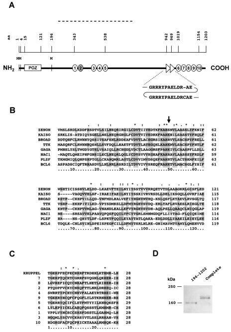
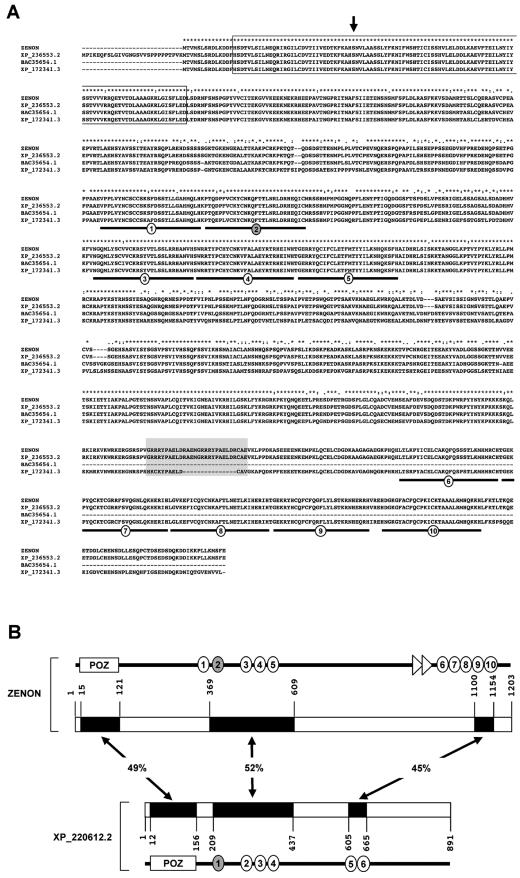
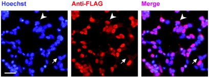
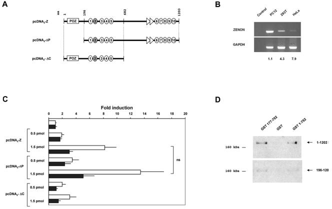
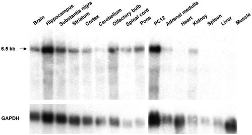
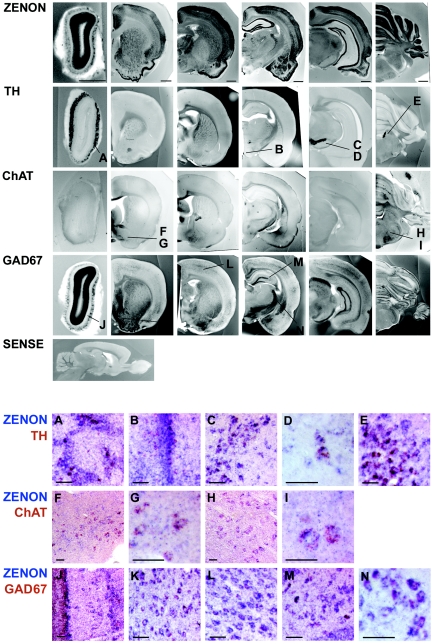
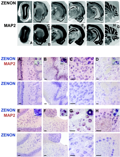
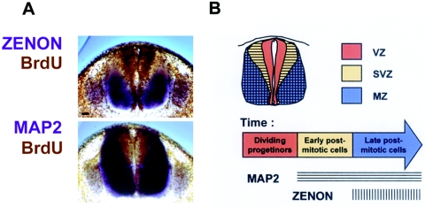
The rat tyrosine hydroxylase gene promoter contains an E-box/dyad motif and an octameric and heptameric element that may be recognized by classes of transcription factors highly expressed during nervous system development. In a one-hybrid genetic screen, we used these sites as targets to isolate cDNAs encoding new transcription factors present in the brain. We identified ZENON, a novel rat POZ protein that contains two clusters of Kruppel-like zinc fingers and that presents several features of a transcription factor. ZENON is found in nuclei following transient transfection with the cDNA. The N-terminal zinc finger cluster contains a DNA binding domain that interacts with the E box. Cotranfection experiments revealed that ZENON induces tyrosine hydroxylase promoter activity. Unlike other POZ proteins, the ZENON POZ domain is not required for either activation of transcription or self-association. In the embryonic neural tube, ZENON expression is restricted to neurons that have already achieved mitosis and are engaged in late stages of neuronal differentiation (late postmitotic neurons). ZENON neuronal expression persists in the adult brain; therefore, ZENON can be considered a marker of mature neurons. We propose that ZENON is involved in the maintenance of panneuronal features and/or in the survival of mature neurons.


Figures

FIG. 1.
FIG. 1.
ZENON is a POZ Kruppel-like zinc finger protein. (A) Schematic diagram of the primary structure of the ZENON protein. The position of each element is indicated by the amino acid (aa) scale above the structure. The fragment isolated by one-hybrid screening is indicated by a broken line. Amino acids are numbered from the most amino-terminal methionine in frame with the POZ domain. Zinc finger motifs are represented as circles numbered from 1 to 10. Zinc finger 2 is a C2HC zinc finger (shaded circle); the others are C2H2 zinc fingers (white circles). A repeat, GRRRYPAELDR-AE/GRRRYPAELDRCAE, is indicated by two triangles. M, methionine. (B) Amino acid alignment of ZENON and other POZ domains. The alignment was generated with ClustalX software. The most highly conserved amino acids are shaded. Asterisks indicate positions at which 100% identity among all of the sequences is observed. Dots indicate positions at which the amino acids are functionally similar. The arrow indicates a position at which K or R is usually found but is not conserved in the ZENON primary sequence. (C) Amino acid sequence alignment of ZENON zinc fingers 1 to 10 with one Kruppel zinc finger. Note the HX4H spacing in zinc finger 5, with X representing any amino acid. This spacing is thought to provide greater flexibility for DNA binding than the more common HX3H spacing. (D) Western blot analysis of the ZENON translation start site in mammalian cells. 293T cells were transfected with a complete (amino acids 1 to 1202) or a partial (amino acids 196 to 1202) ZENON ORF in frame with a 3′-terminal FLAG sequence. The partial construct includes a methionine surrounded by a consensus Kozak sequence (position 196 in panel A). After separation on a polyacrylamide gel, the ZENON-FLAG fusion proteins were detected with an anti-FLAG antibody. The fusion protein obtained with the complete ZENON coding sequence has a higher molecular mass than the protein encoded by the partial sequence, suggesting that translation can start from methionine residues located upstream from the POZ domain (positions 1 and 4 in panel A), despite the absence of a Kozak sequence.
FIG. 2.
FIG. 2.
Amino acid alignment of ZENON with putative polypeptides present in databases. (A) Alignment of ZENON with putative orthologs. XP_236553.2 and XP_172341.2 were annotated from rat and human genomic sequences, respectively. The BAC35654.1 sequence was predicted from a murine cDNA. The alignment was generated with ClustalX software. Asterisks indicate positions at which 100% identity among all of the sequences is observed. Dots indicate positions at which amino acids are functionally similar. The arrow indicates a position at which K or R is usually found among POZ proteins but is not conserved in the primary sequences of ZENON and its orthologs. The POZ domain (boxed sequence), zinc fingers (underlined sequence), and repeat (shaded sequence) are indicated. Note that the repeat is not evolutionarily conserved because it is not present in humans. (B) Schematic alignment of ZENON with a potential homologous rat polypeptide, XP_220612.2. The related regions are indicated by black boxes, and the percentages of amino acids identical between each region of ZENON and its homolog are indicated.
FIG. 3.
FIG. 3.
The ZENON protein is targeted to nuclei. 293T cells were transfected with the complete ZENON coding region (amino acids 1 to 1202) in frame with the FLAG sequence. The ZENON-FLAG fusion protein was detected with an anti-FLAG antibody 48 h after transfection. Nuclei were counterstained with Hoechst reagent. The arrowhead indicates a nucleus containing the ZENON-FLAG fusion protein. The arrow indicates a nucleus in which the ZENON-FLAG fusion protein is not present, probably because the cell was not transfected. Bar, 20 μm.
FIG. 4.
FIG. 4.
Interaction of ZENON zinc finger domains with the E box. (A) Sodium dodecyl sulfate-polyacrylamide gel electrophoresis of purified L-ZENON and R-ZENON proteins. L-ZENON was generated from a sequence containing zinc fingers 1 to 5 (left hand), whereas R-ZENON was produced from a sequence containing zinc fingers 6 to 10 (right hand). Approximately 20 μg of L-ZENON and 5 μg of R-ZENON were run on a 9% polyacrylamide gel and stained with Coomassie blue. The L-ZENON sequence gave rise to two products of equal intensities, one full length and one truncated. Both contained the complete left hand of zinc fingers, as indicated by their size. (B) EMSA with a 32P-labeled pTH probe spanning the TH proximal promoter. The pTH probe is shown alone (first lane) or in the presence of 1 μg each of L-ZENON and R-ZENON recombinant proteins (following lanes). Arrows indicate free probe and complexes I and II observed with L-ZENON. (C) EMSA with a 32P-labeled E probe covering the E box and surrounding dyad element. The E probe is shown alone (first lane) or with 1 μg of L-ZENON alone (second lane) or in the presence of orthophenanthroline (o-phenanth; following lanes). (D) EMSA with the 32P-labeled E probe incubated with 1 μg of L-ZENON alone or in the presence of various competitors (molar excess, 50- or 200-fold). Complex intensity represents the sum of complex I and complex II intensities quantified with Gelanalyst software. The results shown are representative of four independent gel retardation assays.
FIG. 5.
FIG. 5.
ZENON modulates TH transcriptional activity independently of the POZ domain. (A) ZENON effectors used in cotransfection experiments. The indicated fragments of the ZENON ORF were introduced into pcDNA3 downstream from a cytomegalovirus promoter to generate pcDNA3-Z, pcDNA3-ΔP, and pcDNA3-ΔC. (B) Comparative RT-PCR analyses of ZENON expression in PC12, 293T, and HeLa cells. The first lane shows a negative control (PC12 cells in the absence of reverse transcriptase). Amplification of GAPDH was used to normalize for RT efficiency in each cell line. Values (induction factors) represent the induction of wild-type TH promoter activity (5kbTH-Luc) by 1 pmol of pcDNA3-Z in each cell line relative to the control (1 pmol of empty pcDNA3). (C) Cotransfection experiments with ZENON effectors in HeLa cells. Reporter plasmids contained the luciferase gene driven by the rat TH promoter, either intact (5kbTH-Luc; white bars) or bearing a mutated E box [5kbTH(ΔE)-Luc; black bars]. Cells were transfected with various amounts of ZENON effector constructs (0 to 1.5 pmol), the total amount of DNA being brought to 1.5 pmol by the addition of insert-free pcDNA3. In the absence of ZENON effectors, the transcriptional activity of 5kbTH-Luc was approximately twice that of 5kbTH(ΔE)-Luc; results are shown as induction of TH promoter activity to facilitate interpretation. Values represent the means ± 95% confidence intervals for five independent experiments performed in triplicate with various plasmid preparations. 5kbTH-Luc induction by ZENON effectors was significant compared to that obtained with empty pcDNA3 (P < 10−4). For each ZENON effector, 5kbTH-Luc induction and 5kbTH(ΔE)-Luc induction were significantly different (P = 0.001). 5kbTH-Luc induction by pcDNA3-ΔC was significantly lower than 5kbTH-Luc induction by pcDNA3-Z and pcDNA3-ΔP (P < 10−5). ns, no significant difference in the induction of TH promoter activity was obtained with pcDNA3-Z and pcDNA3-ΔP (P > 0.01). (D) GST pull-down assay with GST-ZENON fusion proteins GST 177-752 and GST 1-752 and GST alone. GST 177-752 does not include the POZ domain. The fusion proteins and GST were incubated with cell extracts containing the complete or partial ZENON-FLAG polypeptide and then were subjected to Western blot analysis with an anti-FLAG antibody. (Upper panel) Complete ZENON-FLAG polypeptide (1 to 1202). (Lower panel) ZENON-FLAG polypeptide lacking the POZ domain (196 to 1202).
FIG. 6.
FIG. 6.
Northern blot analysis showing the tissue distributions of ZENON mRNA. (Upper panel) Poly(A)+ RNAs from various rat tissues (0.5 to 2 μg/lane) hybridized with a ZENON riboprobe. The arrow indicates the estimated size of ZENON mRNA. (Lower panel) Hybridization with a GAPDH probe to normalize for loading differences.
FIG. 7.
FIG. 7.
ZENON mRNA is present in various neuronal subtypes in adult brain. (Upper panels) Adult rat sections (250 μm) were hybridized with ZENON, TH, ChAT, GAD67, and sense riboprobes, which were detected by NBT-BCIP. Bars, 1 mm. The letters indicate the regions from which doubly stained sections A to N were taken. (Lower panels) Adult rat brain sections (14 μm) were hybridized with Dig- and Fluo-labeled riboprobes, which were consecutively detected by incubation with NBT-BCIP (blue) and INT-BCIP (red) substrates. Bars, 50 μm. (A to E) Dig-ZENON and Fluo-TH riboprobes. A, olfactory bulb; B, hypothalamus; C and D, substantia nigra; E, locus coeruleus. (F to I) Dig-ZENON and Fluo-ChAT riboprobes. F and G, diagonal band; H and I, pons. (J to N) Dig-ZENON and Fluo-GAD67 riboprobes. J, olfactory bulb; K, septum; L, cerebral cortex; M, hippocampus; N, reticulate nucleus of thalamus.
FIG. 8.
FIG. 8.
ZENON mRNA colocalizes with MAP2 mRNA in adult brain. (Upper panels) Serial adult rat sections (250 μm) were hybridized with Dig-ZENON and Dig-MAP2 riboprobes, which were detected by NBT-BCIP. Bars, 1 mm. The letters indicate the regions from which doubly stained sections A to H were taken. (Lower panels) Adult rat brain sections (14 μm) were hybridized with Dig-ZENON and Fluo-MAP2 riboprobes, which were consecutively detected by incubation with NBT-BCIP (blue) and INT-BCIP (red) substrates. For each brain region examined, a double-labeling experiment and in situ hybridization with the Dig-ZENON riboprobe only performed on adjacent sections are shown in parallel. Bars, 50 μm. Insets show higher magnifications (bars, 10 μm) of the doubly labeled cells. A, olfactory bulb; B, pyriform cortex; C, cerebral cortex; D, striatum; E, hippocampus; F, thalamus; G, pons; H, cerebellum.
FIG. 9.
FIG. 9.
ZENON gene expression during development. Rat sections (250 μm) were hybridized with Dig-ZENON and Dig-MAP2 riboprobes, which were detected by NBT-BCIP. Bars, 1 mm. E12 (A to C), E13 (D to F), and E14 (G to I) panels show whole-embryo sections at the E12, E13, and E14 developmental stages, respectively. The plan and level of each section are indicated on the embryo schemes. The arrow indicates ZENON gene expression in the ventral spinal cord. The arrowhead indicates ZENON gene expression in the lateral telencephalon and diencephalon. Asterisks indicate expression in the trigeminal and dorsal root ganglia. E17 (J to K) and P1 (L to N) panels show coronal hemisections of the forebrain and hindbrain at the E17 and P1 developmental stages, respectively.
FIG. 10.
FIG. 10.
ZENON is a marker of late postmitotic neurons in the developing spinal cord. (A) E14 rat sections (250 μm) were hybridized with Dig-ZENON and Dig-MAP2 riboprobes, which were detected by NBT-BCIP. Samples were subsequently subjected to immunohistochemical analysis to determine BrdU incorporation. Bar, 50 μm. (B) Interpretation correlating ZENON and MAP2 expression patterns with the timing of differentiation. VZ, ventricular zone; SVZ, subventricular zone; MZ, marginal zone.

Similar articles

Cited by

References

    1. Adhikary, S., K. Peukert, H. Karsunky, V. Beuger, W. Lutz, H. P. Elsasser, T. Moroy, and M. Eilers. 2003. Miz1 is required for early embryonic development during gastrulation. Mol. Cell. Biol. 23:7648-7657. - PMC - PubMed
    1. Albagli, O., P. Dhordain, C. Deweindt, G. Lecocq, and D. Leprince. 1995. The BTB/POZ domain: a new protein-protein interaction motif common to DNA- and actin-binding proteins. Cell Growth Differ. 6:1193-1198. - PubMed
    1. Albagli-Curiel, O. 2003. Ambivalent role of BCL6 in cell survival and transformation. Oncogene 22:507-516. - PubMed
    1. Albanèse, V., N. F. Biguet, H. Kiefer, E. Bayard, J. Mallet, and R. Meloni. 2001. Quantitative effects on gene silencing by allelic variation at a tetranucleotide microsatellite. Hum. Mol. Genet. 10:1785-1792. - PubMed
    1. Altschul, S. F., W. Gish, W. Miller, E. W. Myers, and D. J. Lipman. 1990. Basic local alignment search tool. J. Mol. Biol. 215:403-410. - PubMed

Publication types

MeSH terms

LinkOut - more resources