. 2005 Mar;25(5):1779-92.
doi: 10.1128/MCB.25.5.1779-1792.2005.

An acidic protein, YBAP1, mediates the release of YB-1 from mRNA and relieves the translational repression activity of YB-1

Affiliations

An acidic protein, YBAP1, mediates the release of YB-1 from mRNA and relieves the translational repression activity of YB-1

Ken Matsumoto et al. Mol Cell Biol. 2005 Mar.

Abstract

Eukaryotic Y-box proteins are nucleic acid-binding proteins implicated in a wide range of gene regulatory mechanisms. They contain the cold shock domain, which is a nucleic acid-binding structure also found in bacterial cold shock proteins. The Y-box protein YB-1 is known to be a core component of messenger ribonucleoprotein particles (mRNPs) in the cytoplasm. Here we disrupted the YB-1 gene in chicken DT40 cells. Through the immunoprecipitation of an epitope-tagged YB-1 protein, which complemented the slow-growth phenotype of YB-1-depleted cells, we isolated YB-1-associated complexes that likely represented general mRNPs in somatic cells. RNase treatment prior to immunoprecipitation led to the identification of a Y-box protein-associated acidic protein (YBAP1). The specific association of YB-1 with YBAP1 resulted in the release of YB-1 from reconstituted YB-1-mRNA complexes, thereby reducing the translational repression caused by YB-1 in the in vitro system. Our data suggest that YBAP1 induces the remodeling of YB-1-mRNA complexes.


Figures

FIG. 1.
FIG. 1.
Generation of a YB-1/ clone. (A) Schematic representation of part of the YB-1 locus, the gene disruption constructs, and the configuration of the targeted loci. Black boxes indicate the positions of the exons. RI, EcoRI recognition sites. The sizes of the hybridized fragments of the wild-type and targeted loci in Southern blotting (B) are shown. (B) Southern blot analysis of EcoRI-digested genomic DNA from the indicated genotypes with the probe shown in panel A. (C) Immunoblot analysis of total cell lysates with anti-YB-1 antibodies. The positions of molecular mass markers are shown on the left. (D) Growth curves for DT40 cells and YB-1 gene disruptants. DT40 cells (circles), YB-1+/ cells (triangles), and YB-1/ cells (squares), which had been maintained at 39.5°C, were inoculated at 5 × 10⁴ cells/ml and incubated at 39.5°C (left panel) or 33°C (right panel). The cell numbers were determined at the time points indicated. (E) Protein expression after the temperature shift. DT40 and YB-1/ cells were cultured at 39.5 or 33°C for the times indicated and harvested, and 5 μg of total protein from the cell lysates was analyzed by immunoblotting with anti-YB-1 and anti-α-tubulin antibodies.
FIG. 2.
FIG. 2.
Complementation of the growth defects of the YB-1 gene disruptants by FLAG-tagged YB-1 expression. (A) Lysates of DT40, YB-1/, and YB-1//YB-1f cells were examined for the expression of YB-1 by immunoblotting with anti-FLAG (top left panel) or anti-YB-1 (top right panel) antibodies. The same membranes were reprobed with anti-HuR antibodies (bottom panels). (B) Growth curves. DT40 cells (circles) and YB-1//YB-1f cells (triangles), which had been maintained at 39.5°C, were inoculated at 5 × 10⁴ cells/ml and incubated at 39.5°C (open symbols) or 33°C (closed symbols). The cell numbers were determined at the time points indicated. (C) DT40 and YB-1//YB-1f cell lysates were fractionated with sucrose gradients in the presence of EDTA. Fractions were collected from the top of the gradients. Proteins from each fraction were analyzed by immunoblotting with anti-YB-1 (DT40) and anti-FLAG (YB-1//YB-1f) antibodies. (Top panel) Absorbance profile at 254 nm for the gradient of DT40 cell lysates. (Bottom panel) Gel electrophoresis pattern of total RNA from each fraction. The positions of 28S and 18S rRNAs are indicated; 40S and 60S indicate the positions of ribosomal subunits.
FIG. 3.
FIG. 3.
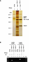
Isolation of YB-1-associated complexes. (A) DT40 cell lysates (lane 1) and YB-1//YB-1f cell lysates before (lane 2) or after (lane 3) RNase A treatment were incubated with anti-FLAG antibody beads, and bound proteins were eluted as described in Materials and Methods. Eluted proteins were resolved by SDS-PAGE and detected by silver staining. (B) RNA in immunoprecipitates obtained with anti-FLAG antibodies was used as the template for RT-PCR to detect chicken β-actin mRNA. In lanes 4 and 8, reverse transcriptase was omitted during RT-PCR. 3′UTR, 3′ untranslated region. Lane M, markers.
FIG. 4.
FIG. 4.
Interaction between YB-1 and YBAP1. (A) Coimmunoprecipitation of chicken YBAP1 with YB-1. Lysates of DT40 (lane 1) and YB-1//YB-1f (lane 2) cells and immunoprecipitates (IP) obtained with anti-FLAG antibodies from DT40 and YB-1//YB-1f cell lysates before or after RNase A treatment (lanes 3 to 5) (Fig. 3A shows silver staining of total protein) were examined by immunoblotting with anti-38K protein/YBAP1 antibodies. (B) GST pull-down assay. Recombinant YBAP1 was incubated with GST (lane 1) or GST-YB-1 (lane 2) bound to glutathione-Sepharose. The bound materials were analyzed by immunoblotting with anti-38K protein/YBAP1 antibodies. An aliquot of recombinant YBAP1 was analyzed in parallel (lane 3). (C) Domain of YB-1 required for interaction with YBAP1. Full-length YB-1 (wild type [WT]) and deletion mutants of YB-1 with a C-terminal FLAG tag were generated in an in vitro transcription-translation system. Each reaction mixture was incubated with GST or GST-YBAP1 bound to glutathione-Sepharose. The bound materials were analyzed by immunoblotting with anti-FLAG antibodies. Schematic diagrams of the constructs are shown. (D) Sucrose gradient fractionation of DT40 cell lysates in the presence of cycloheximide. Proteins from each fraction were analyzed by immunoblotting with anti-38K protein/YBAP1 and anti-YB-1 antibodies. (Top panel) Absorbance profile at 254 nm. (Bottom panel) Gel electrophoresis pattern of total RNA from each fraction. The positions of 28S and 18S rRNAs are indicated.
FIG. 5.
FIG. 5.
YBAP1 remodels YB-1-mRNPs. (A) One microgram each of purified His6-YB-1 and His6-YBAP1 was analyzed by SDS-PAGE and stained with Coomassie brilliant blue. The sizes of the molecular mass markers (Bio-Rad) are shown on the left. (B) Gel retardation assay of reconstituted YB-1-mRNPs. One picomole of 32P-labeled β-actin mRNA and 0 μg (lane 1), 0.35 μg (10 pmol; lane 2), 0.7 μg (20 pmol; lane 3), 1.4 μg (40 pmol; lane 4), 2.1 μg (60 pmol; lane 5), and 2.8 μg (80 pmol; lane 6) of recombinant His6-YB-1 were incubated and electrophoresed in an agarose gel. (C) His6-YB-1 and His6-YBAP1 were incubated, and then β-actin mRNA was added to the reaction mixtures as described in Materials and Methods. The mixtures were electrophoresed in an agarose gel.
FIG. 6.
FIG. 6.
YBAP1 relieves translational repression mediated by YB-1. (A) Translational repression by YB-1. 32P-labeled β-actin mRNA and the indicated amounts of His6-YB-1 were incubated with wheat germ extracts in the presence of [35S]methionine. Translational products were analyzed by SDS-PAGE and quantified. The graph shows relative [35S]methionine incorporation into synthesized β-actin. The values were normalized to the value in lane 1, which was 100%. After the translation reaction, β-actin mRNA was recovered and analyzed in an agarose gel containing formaldehyde. Lane C shows an aliquot of the template RNA. (B) β-actin mRNA was incubated with YB-1-YBAP1 complexes and wheat germ extracts. Translational products and RNA were analyzed as described for panel A.
FIG. 7.
FIG. 7.
YBAP1 relieves translational repression of CAT mRNA mediated by YB-1. (A) His6-YB-1 and His6-YBAP1 were incubated, and then 1 pmol of 32P-labeled CAT mRNA was added to the reaction mixtures as described in Materials and Methods. The mixtures were electrophoresed in an agarose gel. (B) CAT mRNA was incubated with YB-1-YBAP1 complexes and wheat germ extracts. Translational products and RNA were analyzed as described in the legend to Fig. 6A.
FIG. 8.
FIG. 8.
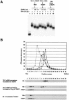
YBAP1 displaces YB-1 molecules from YB-1-mRNPs. (A) YBAP1 remodels preformed YB-1-mRNPs. Lane 1, naked 32P-labeled β-actin mRNA. Lanes 2 to 4, His6-YB-1 (1.4 μg) was preincubated for 15 min at 30°C with the indicated amounts of His6-YBAP1. β-actin mRNA then was added, and the mixtures were incubated for an additional 20 min at 30°C. Lanes 5 to 7, His6-YB-1 was preincubated for 15 min at 30°C with β-actin mRNA. His6-YBAP1 then was added, and the mixtures were incubated for an additional 20 min at 30°C. Lanes 8 to 10, His6-YB-1, His6-YBAP1, and β-actin mRNA were mixed and incubated for 20 min at 30°C. Aliquots of the mixtures were analyzed in an agarose gel. (B) YB-1-mRNPs were prepared by incubating 20 pmol of 32P-labeled β-actin mRNA and 42 μg of His6-YB-1 for 20 min at 30°C and purified through a sucrose gradient (data not shown). Purified YB-1-mRNPs containing 3 pmol of mRNA were incubated for 20 min at 30°C without His6-YBAP1 (closed squares) or with 0.9 μg (closed triangles) or 3 μg (closed circles) of His6-YBAP1. The mixtures were centrifuged in 15 to 40% sucrose gradients at 50,000 rpm for 4 h in an SW55 Ti rotor (Beckman). Two control reactions, (i) His6-YB-1 and His6-YBAP1 incubated without RNA and (ii) naked β-actin mRNA (open circles), were analyzed in parallel. (Top panel) The gradient was collected into 20 fractions from the top, and the radioactivity in each fraction was quantified. (Bottom panels) YB-1 and YBAP1 in each fraction were analyzed by immunoblotting.

Similar articles

Cited by

References

    1. Akey, C. W., and K. Luger. 2003. Histone chaperones and nucleosome assembly. Curr. Opin. Struct. Biol. 13:6-14. - PubMed
    1. Aoki, K., K. Matsumoto, and M. Tsujimoto. 2003. Xenopus cold-inducible RNA-binding protein 2 interacts with ElrA, the Xenopus homolog of HuR, and inhibits deadenylation of specific mRNAs. J. Biol. Chem. 278:48491-48497. - PubMed
    1. Bae, W., P. G. Jones, and M. Inouye. 1997. CspA, the major cold shock protein of Escherichia coli, negatively regulates its own gene expression. J. Bacteriol. 179:7081-7088. - PMC - PubMed
    1. Baer, B. W., and R. D. Kornberg. 1983. The protein responsible for the repeating structure of cytoplasmic poly(A)-ribonucleoprotein. J. Cell Biol. 96:717-721. - PMC - PubMed
    1. Bargou, R. C., K. Jurchott, C. Wagener, S. Bergmann, S. Metzner, K. Bommert, M. Y. Mapara, K. J. Winzer, M. Dietel, B. Dorken, and H. D. Royer. 1997. Nuclear localization and increased levels of transcription factor YB-1 in primary human breast cancers are associated with intrinsic MDR1 gene expression. Nat. Med. 3:447-450. - PubMed

Publication types

MeSH terms

LinkOut - more resources