. 2005 Mar;25(5):1793-803.
doi: 10.1128/MCB.25.5.1793-1803.2005.

Mitogen-activated protein kinases with distinct requirements for Ste5 scaffolding influence signaling specificity in Saccharomyces cerevisiae

Affiliations

Mitogen-activated protein kinases with distinct requirements for Ste5 scaffolding influence signaling specificity in Saccharomyces cerevisiae

Laura J Flatauer et al. Mol Cell Biol. 2005 Mar.

Abstract

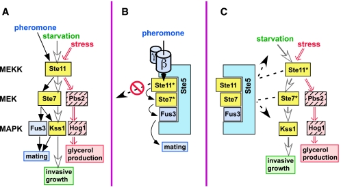
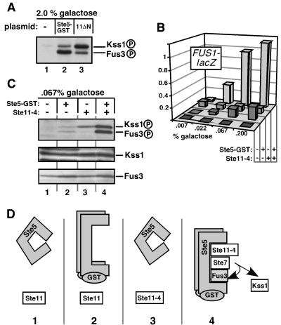
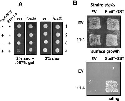
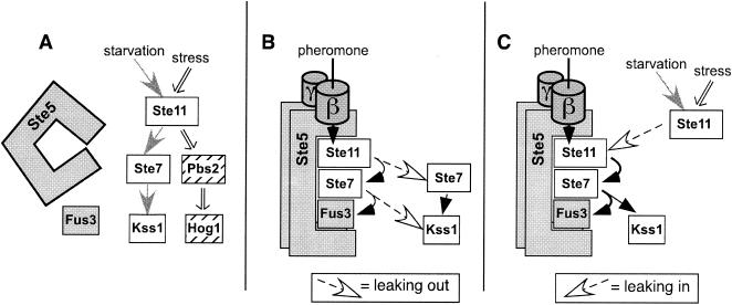
Scaffold proteins are believed to enhance specificity in cell signaling when different pathways share common components. The prototype scaffold Ste5 binds to multiple components of the Saccharomyces cerevisiae mating pheromone response pathway, thereby conducting the mating signal to the Fus3 mitogen-activated protein kinase (MAPK). Some of the kinases that Ste5 binds to, however, are also shared with other pathways. Thus, it has been presumed that Ste5 prevents its bound kinases from transgressing into other pathways and protects them from intrusions from those pathways. Here we found that Fus3MAPK required Ste5 scaffolding to receive legitimate signals from the mating pathway as well as misdirected signals leaking from other pathways. Furthermore, increasing the cellular concentration of active Ste5 enhanced the channeling of inappropriate stimuli to Fus3. This aberrant signal crossover resulted in the erroneous induction of cell cycle arrest and mating. In contrast to Fus3, the Kss1 MAPK did not require Ste5 scaffolding to receive either authentic or leaking signals. Furthermore, the Ste11 kinase, once activated via Ste5, was able to signal to Kss1 independently of Ste5 scaffolding. These results argue that Ste5 does not act as a barrier that actively prevents signal crossover to Fus3 and that Ste5 may not effectively sequester its activated kinases away from other pathways. Rather, we suggest that specificity in this network is promoted by the selective activation of Ste5 and the distinct requirements of the MAPKs for Ste5 scaffolding.


Figures

FIG. 1.
FIG. 1.
(A) Shared MAPK cascade components signal to three distinct endpoints. See the text for details. Shared components (Ste11MEKK, Ste7MEK, and Kss1MAPK) are shown in yellow. Nutrient limitation (starvation) is known to activate the invasive growth program, but whether this or some other signal is transmitted by the MAPK cascade is unknown. Whatever this signal is, it appears to be relayed by the Msb2 protein (12). (B and C) Models for how the Ste5 scaffold protein may promote signaling specificity. The active, phosphorylated Ste11 and Ste7 isoforms are indicated by an asterisk. (B) By the selective recognition of mating components, Ste5 enhances signaling within the mating pathway. Also, Ste5 may sequester its bound components and prevent them from straying into other pathways. (C) Ste5 may act as a sequestering barrier to protect mating components, particularly Fus3MAPK, from Ste11MEKK or Ste7MEK activated by other pathways.
FIG. 2.
FIG. 2.
Fus3MAPK activation during mating requires Ste5 scaffolding, but Kss1MAPK activation does not. (A) Strains containing wild-type Ste5 (WT), lacking Ste5 (ste5Δ), or containing the adapter-defective mutant Ste5F514L (F514L) or the scaffolding-defective mutant Ste5V763A S861P (VASP) were treated (+) or not (−) with 1 μM α-factor mating pheromone (phm) for 15 min. Activation loop phosphorylation was detected by immunoblotting with a phosphorylation state-specific antibody (αpTEpY). Kss1-P and Fus3-P indicate the dually phosphorylated, activated species. Total protein levels were determined with anti-Kss1 (αKss1) and anti-Fus3 (αFus3) antisera. (B) Schematic interpretation of the results; see the text for details.
FIG. 3.
FIG. 3.
Differential requirements of the Fus3 and Kss1 MAPKs for Ste5 scaffolding during invasive growth and the osmotic stress response. (A) Fus3MAPK activation during invasive growth requires Ste5, but Kss1MAPK activation does not. Strains containing wild-type Ste5 (WT), lacking Ste5 (ste5Δ), or lacking Ste5 and Ste7 (ste5Δ ste7Δ) were grown on plates for 24 h and assayed for MAPK phosphorylation. FRE-lacZ reporter gene activity was also measured; standard error bars are shown (n = 7). Results were normalized by setting the mean β-galactosidase activity of the wild-type strain (1,200 U/min/mg of total yeast protein) to 1.0. Similar results were obtained when cells were grown in liquid prior to harvesting for analysis of FRE-lacZ expression (data not shown). Parallel plates were washed and scored for invasive growth. (B) Concurrent leaks. The invasive growth pathway contributes to the basal phosphorylation of Fus3, and the mating pathway contributes to the basal phosphorylation of Kss1. Strains containing wild-type Ste4 and/or Msb2 (+) or lacking Ste4 and/or Msb2 (Δ) were grown in liquid to mid-log phase and assayed for MAPK phosphorylation. Similar results were obtained when the cells were grown on plates for 24 h (data not shown). FUS1-lacZ and FRE-lacZ reporter gene activity was also measured; error bars represent standard deviations (n = 2). Note that the FUS1-lacZ expression being measured is the basal level (i.e., in the absence of pheromone stimulation). (C) Leaking from the osmostress pathway is preferentially channeled into Kss1, particularly when Ste5 is absent. Liquid cultures of strains containing wild-type Ste5 (STE5+ pbs2Δ) or not (ste5Δ pbs2Δ) were treated with 0.7 M sorbitol for 3 h after reaching mid-log phase. Lysates were probed for MAPK phosphorylation, and FUS1-lacZ or FRE-lacZ reporter gene activity was measured. The β-galactosidase activity for the reference strain (normalized to 1.0) was 1,200 U/min/mg of protein (FUS1-lacZ) and 1,150 U/min/mg of protein (FRE-lacZ). Error bars represent standard deviations (n = 2).
FIG. 4.
FIG. 4.
Influence of Ste5 on Ste11-4-generated signaling to Kss1MAPK and Fus3MAPK. + indicates the wild-type allele, Δ indicates a gene deletion, and 11-4 indicates strains containing Ste11-4. (A) Ste11-4-stimulated phosphorylation of Fus3 but not of Kss1 requires Ste5. Cells were grown on plates for 24 h before harvesting. The immunoblot in this figure was exposed for a shorter time than the immunoblot in Fig. 3A; this is why the equivalent lane 1's look different. (B) FRE-lacZ expression in the strains shown in A (upper panel) and FUS1-lacZ expression of strains grown in liquid and treated (+) or not (−) with 1 μM pheromone for 2 h (lower panel). Standard deviations are shown (n = 2 to 4). (C) Scaffolding-defective mutant of Ste5 (V763A S861P) does not support Ste11-4-stimulated Fus3 phosphorylation. (D) Removal of endogenous Ste11 has no effect on the inability of plasmid-expressed Ste11-4 to efficiently stimulate Fus3 phosphorylation. Cells were grown on plates for 24 h before harvesting. (E) Effect of Kss1 removal and pheromone stimulation on Fus3 phosphorylation in STE11-4 strains. Liquid-grown cells were treated or not for 15 min with 1 μM pheromone.
FIG. 5.
FIG. 5.
Active Ste5 guides signals carried by Ste11MEKK to Fus3MAPK during Ste11-4-generated signaling. Ste5-GST expression was regulated by galactose; the percent galactose used is shown. (A) MAPK phosphorylation by overexpressed and constitutively active Ste5 or Ste11. Ste11ΔN expression (lane 3) was also controlled by galactose levels. (B) FUS1-lacZ reporter gene expression in strains grown on plates containing 2% sucrose and the inducer galactose at increasing concentrations (0.007% to 0.2%). Strains containing Ste5-GST or Ste11-4 are indicated with a + in the appropriate row. (C) MAPK activation loop phosphorylation of the strains from B induced by 2% sucrose and 0.067% galactose. (D) Illustrative description and interpretation of the experiment in C. Panels 1 to 4 correspond to lanes 1 to 4 in C. Active Ste5 has no signal to channel (panel 2), and Ste11-4 needs active Ste5 to hyperactivate Fus3 (panel 3). Together, active Ste5 can channel the Ste11-4 signal to Fus3 (panel 4).
FIG. 6.
FIG. 6.
Ste5-promoted signal crossover results in miscued induction of cell cycle arrest and mating. (A) Cell cycle arrest in strains with active Ste5 and Ste11-4. Wild-type (WT) and fus3Δ strains were transformed with Ste5-GST and/or Ste11-4 or the corresponding empty vector as indicated, and low-level Ste5-GST expression was induced or not by growth on plates containing 2% sucrose and 0.067% galactose or 2% dextrose, respectively. Equal numbers of cells (roughly 30,000) were spotted and grown for 6 days. (B) Mating in strains with active Ste5 and Ste11-4. MATa strains lacking Ste4 and containing Ste5-GSTC177A,C180A (Ste5*-GST) and/or Ste11-4 or the corresponding empty vector (EV) were grown on a plate containing 2% sucrose and 0.067% galactose for 24 h to induce low-level Ste5-GST (surface growth) and then incubated with a MATα strain and transferred to 2% dextrose plates selective for diploids (mating). Ste5-GSTC177A,C180A is a mutant of Ste5-GST that is active even in the absence of Ste4 (23).
FIG. 7.
FIG. 7.
Model for specific signaling by the selective activation of the Ste5 scaffold protein combined with MAPKs with different scaffold requirements. (A) Fus3MAPK requires active Ste5 in order to effectively receive any signals. Hence, in nonmating cells, Fus3 mostly ignores signals from the invasive growth and osmostress pathways because Ste5 is inactive. Kss1MAPK uses an unknown scaffold or is scaffold independent. Hog1MAPK may use Pbs2MEK as a scaffold (37, 50). When inactive, Ste5 is presumably unable to organize Ste11MEKK, Ste7MEK, and Fus3 into a signaling-competent configuration. Here we have pictured this as a lack of Ste5-kinase association (48). (B and C) In mating cells, activated Ste5 relays the pheromone signal via activated Ste11 to Fus3. However, activated Ste11 can apparently also leak out and transmit signals in a scaffold-independent fashion (B). Furthermore, active Ste5 can also accept Ste11 activated by the other pathways and channel those signals to Fus3, promoting leaking (C).

Similar articles

Cited by

References

    1. Andersson, J., D. M. Simpson, M. Qi, Y. Wang, and E. A. Elion. 2004. Differential input by Ste5 scaffold and Msg5 phosphatase route a MAPK cascade to multiple outcomes. EMBO J. 23:2564-2576. - PMC - PubMed
    1. Bardwell, A. J., L. J. Flatauer, K. Matsukuma, J. Thorner, and L. Bardwell. 2001. A conserved docking site in MEKs mediates high-affinity binding to MAP kinases and cooperates with a scaffold protein to enhance signal transmission. J. Biol. Chem. 276:10374-10386. - PMC - PubMed
    1. Bardwell, L. 2005. A walk-through of the yeast mating pheromone response pathway. Peptides 26:339-350. - PMC - PubMed
    1. Bardwell, L., J. G. Cook, E. C. Chang, B. R. Cairns, and J. Thorner. 1996. Signaling in the yeast pheromone response pathway: specific and high-affinity interaction of the mitogen-activated protein (MAP) kinases Kss1 and Fus3 with the upstream MAP kinase kinase Ste7. Mol. Cell. Biol. 16:3637-3650. - PMC - PubMed
    1. Bardwell, L., J. G. Cook, J. X. Zhu-Shimoni, D. Voora, and J. Thorner. 1998. Differential regulation of transcription: repression by unactivated mitogen-activated protein kinase Kss1 requires the Dig1 and Dig2 proteins. Proc. Natl. Acad. Sci. USA 95:15400-15405. - PMC - PubMed

Publication types

MeSH terms

Substances