. 2005 Mar 1;102(9):3225-9.
doi: 10.1073/pnas.0408870102. Epub 2005 Feb 15.

Structure of a mitochondrial supercomplex formed by respiratory-chain complexes I and III

Affiliations

Structure of a mitochondrial supercomplex formed by respiratory-chain complexes I and III

Natalia V Dudkina et al. Proc Natl Acad Sci U S A. .

Abstract

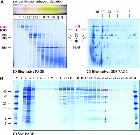
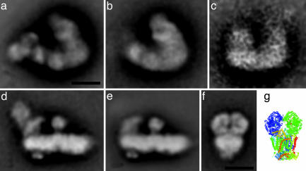
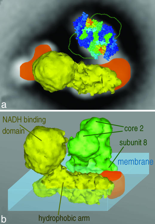
Mitochondria are central to the efficient provision of energy for eukaryotic cells. The oxidative-phosphorylation system of mitochondria consists of a series of five major membrane complexes: NADH-ubiquinone oxidoreductase (commonly known as complex I), succinate-ubiquinone oxidoreductase (complex II), ubiquinol-cytochrome c oxidoreductase (cytochrome bc1 complex or complex III), cytochrome c-O2 oxidoreductase (complex IV), and F1F0-ATP synthase (complex V). Several lines of evidence have recently suggested that complexes I and III-V might interact to form supercomplexes. However, because of their fragility, the structures of these supercomplexes are still unknown. A stable supercomplex consisting of complex I and dimeric complex III was purified from plant mitochondria. Structural characterization by single-particle EM indicates a specific type of interaction between monomeric complex I and dimeric complex III in a 1:1 ratio. We present a model for how complexes I and III are spatially organized within the I+III2 supercomplex.


Figures

Fig. 1.
Fig. 1.
Purification of mitochondrial I+III2 supercomplex, monomeric complex I, and dimeric complex III from A. thaliana. (A) Isolated mitochondria were solubilized by digitonin, and protein complexes were subsequently resolved by sucrose-gradient ultracentrifugation (Upper Left). BN/PAGE was carried out to analyze the protein-complex content of the fractions (Lower Left). Identities of the resolved complexes on the 1D gel were elucidated by a parallel 2D BN/SDS/PAGE of total mitochondrial protein (Right). I+III2, supercomplex formed of complex I and dimeric complex III; I, complex I; H, heat-stress protein 60 complex; V, ATP synthase complex; III2, dimeric complex III; FDH, formate dehydrogenase complex. The molecular masses (in kDa) of standard proteins are given to the left of the 1D gel and above the 2D gel. Fractions 2 and 3 of the sucrose gradient were used for EM analysis of the I+III2 supercomplex, and fraction 4 was used for EM analysis of monomeric complex I. (B) A mitochondrial-membrane fraction was solubilized by 3% Triton X-100, and complex III was subsequently purified by cytochrome-c-affinity chromatography (15). Proteins were eluted from the affinity column by a linear Tris/acetate gradient (20–200 mM) and analyzed by 1D SDS/PAGE (15). Fractions 1–4, flow-through; fractions 5–16, mitochondrial proteins not related to complex III; fractions 17–22, purified complex III (subunits are indicated by arrows). Fraction 21 was used for EM analysis of complex III. The molecular masses (in kDa) of standard proteins are given to the left.
Fig. 2.
Fig. 2.
Projection maps at 18 Å of the Arabidopsis I+III2 supercomplex and its components obtained by single-particle averaging. (a) Averaged projection map of 1,073 top-view projections of I+III2 supercomplex particles, viewed from the matrix side of the membrane. (b) Average of the best 580 top views of a I+III2 supercomplex fragment. (c) Averaged side view of 22 projections of a I+III2 supercomplex fragment. (d) Average of 930 side-view projections of complex I. (e) Average of 2,100 side-view projections of complex I lacking NAD-oxidizing subunits. (f) Average of 1,014 projections of complex III in a side-view position. (g) View of an x-ray structural model of dimeric bovine complex III (24) in a position similar to that in the EM data, with the bulky matrix-exposed domains in the upper part. (Scale bar, 10 nm.)
Fig. 3.
Fig. 3.
Model for the structure of the I+III2 supercomplex from Arabidopsis, incorporating the x-ray structure of complex III and the EM 3D density of complex I from beef heart. (a) Fit within the supercomplex top view of the complex I structure (yellow) and the membrane-embedded lower half of complex III (space-filling structure) seen from the matrix side. The projections of additional complex I densities present in Arabidopsis are in orange, and the outline of the hydrophilic domains of complex III within the matrix is indicated by a green dotted line. (b) Overview of the fitting of complex I and complex III (green), seen from an angle of 45° out of the membrane plane. Components of complexes I and III and the membrane outside the supercomplex (blue) are indicated. Note that the x-ray structure was truncated to 18 Å by using routines from the eman package (41) and displayed with vis5d software (www.ssec.wisc.edu/~billh/vis5d.html).

Similar articles

Cited by

References

    1. Hatefi, Y. (1985) Annu. Rev. Biochem. 54, 1015-1069. - PubMed
    1. Arnold, I., Pfeiffer, K., Neupert, W., Stuart, R. A. & Schägger, H. (1998) EMBO J. 17, 7170-7178. - PMC - PubMed
    1. Schägger, H. & Pfeiffer, K. (2000) EMBO J. 19, 1777-1783. - PMC - PubMed
    1. Schägger, H. & Pfeiffer, K. (2001) J. Biol. Chem. 276, 37861-37867. - PubMed
    1. Eubel, H., Jänsch, L. & Braun, H. P. (2003) Plant Physiol. 133, 274-286. - PMC - PubMed

Publication types

MeSH terms

Substances