. 2005 Feb 15;19(4):502-13.
doi: 10.1101/gad.320705.

Xenopus polo-like kinase Plx1 regulates XErp1, a novel inhibitor of APC/C activity

Affiliations

Xenopus polo-like kinase Plx1 regulates XErp1, a novel inhibitor of APC/C activity

Andreas Schmidt et al. Genes Dev. .

Abstract

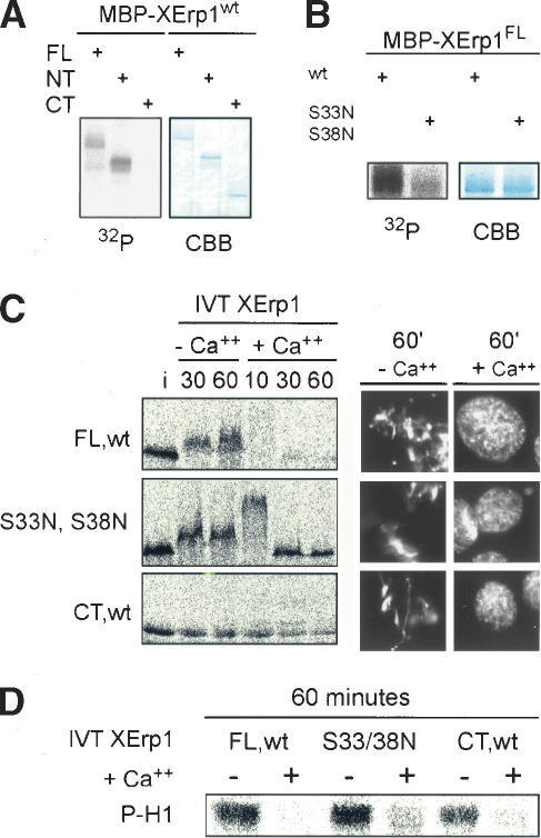
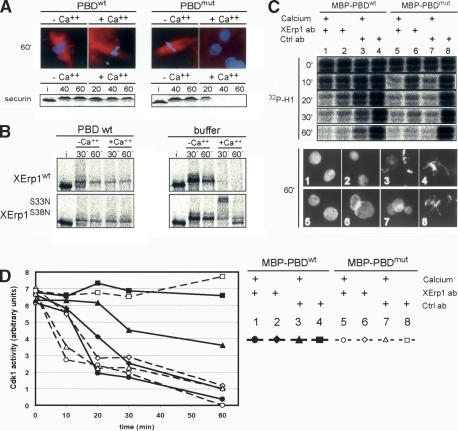
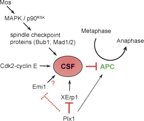
Metaphase-to-anaphase transition is a fundamental step in cell cycle progression where duplicated sister-chromatids segregate to the future daughter cells. The anaphase-promoting complex/cyclosome (APC/C) is a highly regulated ubiquitin-ligase that triggers anaphase onset and mitotic exit by targeting securin and mitotic cyclins for destruction. It was previously shown that the Xenopus polo-like kinase Plx1 is essential to activate APC/C upon release from cytostatic factor (CSF) arrest in Xenopus egg extract. Although the mechanism by which Plx1 regulates APC/C activation remained unclear, the existence of a putative APC/C inhibitor was postulated whose activity would be neutralized by Plx1 upon CSF release. Here we identify XErp1, a novel Plx1-regulated inhibitor of APC/C activity, and we demonstrate that XErp1 is required to prevent anaphase onset in CSF-arrested Xenopus egg extract. Inactivation of XErp1 leads to premature APC/C activation. Conversely, addition of excess XErp1 to Xenopus egg extract prevents APC/C activation. Plx1 phosphorylates XErp1 in vitro at a site that targets XErp1 for degradation upon CSF release. Thus, our data lead to a model of APC/C activation in Xenopus egg extract in which Plx1 targets the APC/C inhibitor XErp1 for degradation.


Figures

Figure 1.
Figure 1.
Domain structure of XErp1 and characterization of the XErp1 antibody. (A) The N terminus of XErp1 contains the putative SCFβ-TRCP recognition sequence DSGX3S. In addition, XErp1 protein contains a putative C-terminal F-box and a zinc-binding region (ZBR). The C terminus of XErp1 shares 39% sequence identity with Xenopus Emi1. (B) ClustalW alignment of XErp1 protein sequences from different vertebrate species shows the evolutionary conservation of the XErp1 proteins. The amino acid sequences of XErp1 peptides that were identified by LC-MS/MS analysis of XErp1 are indicated (solid blue lines, peptide sequences in agreement with both XErp1 and Fbx26 ORF; dashed blue lines, peptide sequences in agreement only with XErp1 ORF). (C) Characterization of XErp1 antibodies. CSF extract or in vitro translated (IVT) XErp1, IVT xEmi1, or unprogrammed wheat germ extract were immunoblotted with affinity purified anti-XErp1 antibodies (left) or with affinity-purified anti-XErp1 antibodies blocked with XErp1 antigen (right). (Middle) The expression level of the 35S-labeled IVT proteins was examined by autoradiographic analysis.
Figure 2.
Figure 2.
XErp1 accumulates during oocyte maturation and is destroyed upon CSF release. (A) CSF-arrested Xenopus egg extract was incubated at 20°C and treated with calcium to induce CSF release in the presence or absence of the translation inhibitor cycloheximide (CHX). At the indicated time points (minutes), samples were taken and immunoblotted for XErp1 protein. In addition, DNA morphology was observed to follow exit from CSF arrest. (B) 35S-labeled IVT XErp1 was immunoprecipitated from CSF-arrested or interphase extract and treated with calf intestinal phosphatase (CIP). (C) Analysis of XErp1 protein levels during oocyte maturation. Stage VI oocytes where isolated and the maturation process induced by addition of progesterone. Oocytes were sampled at the indicated time points (minutes) after progesterone treatment and processed for immunoblot analysis of XErp1 and markers of the maturation process.
Figure 3.
Figure 3.
XErp1 is required for CSF activity. (A) On ice, CSF extracts were treated with immobilized XErp1 antibody preincubated with a 10-fold molar excess of antigen (panel 1), buffer (panel 2), or control antibody preincubated with a 10-fold molar excess of antigen (panel 3). DNA morphology was examined 65 min after warming the extracts to 20°C. Cdk1 activity was determined using an H1 kinase assay from samples withdrawn on ice (0 min) and 10, 20, and 30 min after warming the extracts to 20°C. (B) CSF extracts were incubated for 20 min at 20°C with myc-tagged IVT XErp1 proteins and subsequently incubated with XErp1- or control-antibody beads on ice. Morphology of sperm nuclei and tubulin was examined before depletion and 55 min after warming the XErp1- and mock-depleted samples to 20°C. (C) Total Xenopus extract (T) and the supernatant of depleted extract (SN) were immunoblotted for endogenous XErp1 (middle panel) and for myc-tagged IVT XErp1 proteins (lower panel). The asterisk (*) indicates myc-tagged IVT XErp1FL,wt on the anti-XErp1 immunoblot. 35S-labeled IVT securin was detected by autoradiography. Samples were withdrawn before antibody addition (0) and 30 min after warming the extracts to 20°C (30).
Figure 4.
Figure 4.
Excess XErp1 protein blocks calcium-dependent CSF release independently of Mos/MAPK signaling. (A). Purified MBP-tagged XErp1FL,wt or XErp1FL,C583A was added to Xenopus egg extract at 500 nM final concentration. Chromatin morphology was examined 20 and 60 min after calcium addition. (B) Purified MBP-tagged XErp1FL,wt, XErp1NT,wt, or XErp1CT,wt were separately added to Xenopus egg extract at 100 nM final concentration. Chromatin morphology was examined 30 min and 60 min after calcium addition. (C) Xenopus extract was treated as in A, and samples were taken at the indicated time points after calcium addition and immunoblotted for CDC27. Samples of extract supplemented with 35S-labeled IVT securin or N-terminal fragment of cyclin B1 (cycBNT) were taken at the indicated time points before (CSF) and after calcium addition and analyzed by autoradiography. (D) His-tagged Cdc20 was incubated with buffer (group 2), MBP-XErp1CT,wt (group 3, 300 nM; group 4, 1 μM), or MBP-XErp1NT,wt (group 5, 1 μM; group 6, 3 μM). APC/C immunopurified from mitotic Xenopus extract was then mixed with buffer (group 1) or CDC20 incubated with buffer (group 2) or the indicated MBP-XErp1 fragments (groups 3–6) and assayed for its ability to ubiquitylate 35S-labeled IVT cycBNT at 20°C. At the indicated time points, samples were taken and analyzed by autoradiography. (E) Purified MBP-tagged XErp1FL,wt or control buffer was added to CSF-arrested Xenopus egg extract in the presence or absence of the MAPK kinase inhibitor UO126. Inactivation of the MAPK pathway was confirmed by immunoblotting for active Erk1/2. The cell cycle state of the extract was monitored by the stability of exogenously added 35S-labeled IVT securin.
Figure 5.
Figure 5.
Plx1-dependent destabilization of XErp1 is required for CSF release. (A) MBP-tagged XErp1FL,wt, XErp1NT,wt, and XErp1CT,wt were each incubated in an in vitro phosphorylation reaction with His-tagged Plx1. Incorporation of 32P was analyzed by PAGE and autoradiography. (B) In vitro Plx1 kinase assay as in A using MBP-tagged wild-type XErp1FL,wt or XErp1FL,S33N,S38N. (C) 35S-labeled IVT XErp1FL,wt, XErp1CT,wt, or a mutant form of XErp1 (XErp1FL,S33N,S38N) were incubated in CSF extract (i, input) in the presence or absence of calcium. At the indicated time points, samples were withdrawn and analyzed by autoradiography. (D) Cdk1 activity of all samples shown in C withdrawn after 60 min was measured using an H1 kinase assay.
Figure 6.
Figure 6.
XErp1 is required for effects of dominant-negative PBD on CSF release (A) CSF extracts were incubated with equimolar amounts of MBP-tagged either wild-type (PBDwt) or mutant PBD (PBDmut) of Plx1. Morphology of DNA and tubulin was examined after extracts had been warmed to 20°C for 60 min in the presence or absence of calcium. In addition, the stability of exogenously added 35S-labeled IVT securin was monitored by autoradiography. (B) 35S-labeled IVT XErp1FL,wt or XErp1FL,S33N,S38N were incubated in CSF extract containing PBDwt or control buffer. Extracts were warmed to 20°C, and samples were taken and examined by autoradiography at the indicated time points before and after addition of calcium. (C) Immobilized anti-XErp1 antibodies (lanes 1,2,5,6) or immobilized control antibodies (lanes 3,4,7,8) were added to CSF extract containing PBDWT (lanes 14) or PBDmut (lanes 58) in the presence (lanes 1,3,5,7) or absence (lanes 2,4,6,8) of calcium. Samples were taken at the indicated time points after calcium and/or antibody addition and assayed for Cdk1 activity. In addition, samples of the reactions (panels 18) were withdrawn and chromatin morphology was examined 65 min after warming the extracts to 20°C. (D) Resulting bands of the H1 kinase assay were quantified by densitometry. Band intensities were corrected for background and then plotted against time. Solid lines show reactions with PBDWT; dashed lines show reactions with PBDmut.
Figure 7.
Figure 7.
Model of different pathways regulating CSF activity. The c-Mos/MAPK/p90Rsk pathway seems to exert its inhibitory effect on APC/C by activating components of the spindle assembly checkpoint. The underlying mechanism by which Cdk2/cyclin E inhibits APC/C activity has not been identified. XErp1 and Emi1 mediate CSF activity by inhibiting APC/C directly. Plx1 targets XErp1 and possibly Emi1 for degradation, thereby allowing APC/C activation. Additionally, Plx1 might activate APC/C directly by phosphorylating specific APC/C subunits.

References

    1. Barr F.A., Sillje, H.H., and Nigg, E.A. 2004. Polo-like kinases and the orchestration of cell division. Nat. Rev. Mol. Cell Biol. 5: 429–440. - PubMed
    1. Brassac T., Castro, A., Lorca, T., Le Peuch, C., Doree, M., Labbe, J.C., and Galas, S. 2000. The polo-like kinase Plx1 prevents premature inactivation of the APC(Fizzy)-dependent pathway in the early Xenopus cell cycle. Oncogene 19: 3782–3790. - PubMed
    1. Desai A., Murray, A., Mitchison, T.J., and Walczak, C.E. 1999. The use of Xenopus egg extracts to study mitotic spindle assembly and function in vitro. Methods Cell Biol. 61: 385–412. - PubMed
    1. Descombes P. and Nigg, E.A. 1998. The polo-like kinase Plx1 is required for M phase exit and destruction of mitotic regulators in Xenopus egg extracts. EMBO J. 17: 1328–1335. - PMC - PubMed
    1. Donaldson M.M., Tavares, A.A., Ohkura, H., Deak, P., and Glover, D.M. 2001. Metaphase arrest with centromere separation in polo mutants of Drosophila. J. Cell Biol. 153: 663–676. - PMC - PubMed

Publication types

MeSH terms